A bacteriological study is a study designed to isolate and study their properties in order to make a microbiological diagnosis.
The test material should be taken under aseptic conditions in sterile dishes and delivered to the laboratory as soon as possible. If necessary, samples should be stored refrigerated. The sampling technique depends on the object, the nature of the disease and the properties of the microorganism. One of the most common methods of bacteriological research is bacterioscopy.
To study non-fixed bacteria, two methods are used: crushed (between the slide and coverslip) drop and. It should be remembered that preparations of non-fixed bacteria are contagious.
Smears are used for bacterioscopy of fixed preparations. To prepare them, a drop of the test liquid is spread over the surface of a glass slide and then dried. The most common method of fixing the drug is to carry it through the flame of a gas burner. In some cases, fixing compounds are used. Fixed preparations are usually stained (see Staining of Microorganisms). To the number essential elements bacteriological studies include inoculations and subcultures produced by a bacterial loop or a Pasteur pipette. The loop is sterilized by flame roasting, then cooled by touching an area of uninoculated agar or by rinsing in sterile liquid. When using a Pasteur pipette, its tip is broken off with tweezers, the pipette is carried several times through the flame of the burner and allowed to cool. When sowing, liquid and solid nutrient media are used. When sowing on slant agar, the culture of bacteria is rubbed with a loop over the surface of the agar. When sowing into the thickness of an agar or gelatin column, the nutrient medium is pierced to the bottom of the test tube with a loop or a special needle. When sowing in a liquid medium, it is necessary to ensure that the liquid does not spill out and does not wet the edges of the test tubes and stoppers. Inoculations and subcultures should be carried out near the flame of a gas burner, the test tubes should not remain open for a long time, the loop or Pasteur pipette with the culture should not touch anything; before closing the tube, its edges should be burned. Inoculated tubes must be labeled immediately.
The most important step in bacteriological research is identification - determination of the species or type of bacteria obtained in the form of a pure culture. When identifying bacteria, their physiological and biochemical properties, toxin formation are studied. Serological methods for identifying bacteria are widely used (reactions and). In many cases, the biological method of identification of microorganisms is effective, based on the infection of laboratory animals with the test material or the resulting culture of bacteria and the identification of characteristic pathological changes in animals.
Mechanical and biological methods are used to isolate pure cultures. An example of a mechanical method: a drop of the test material is rubbed with the same sterile spatula or bacterial loop over a dense surface. growth medium, sequentially in the first, second and third . The isolation of a pure culture is made from grown individual colonies and consists in their study and screening on a fresh nutrient medium. Biological methods for isolating pure cultures are based on taking into account one or another property of the isolated microbe, which distinguishes it from other microbes present in the material under study.
In the biological method, this kind of nutrient media is used, in which conditions are created that are favorable for the development of a certain type of microbes. Biological methods also include infection of laboratory animals sensitive to the isolated type of bacteria.
Bacteriological research is a set of methods for detecting pathogenic microorganisms in a patient, in a carrier or on environmental objects. Bacteriological studies They are also used to detect conditionally pathogenic and sanitary-indicative microbes that characterize the degree of pollution of the external environment, to study the microbial landscape of a certain environment (object). Bacteriological research can be used for the diagnosis, prevention of infectious diseases, for the sanitary and hygienic characteristics of the environment surrounding a person, for scientific research.
The material and method of bacteriological research depend on the purpose of the analysis, environmental conditions, pathogenesis and course of the disease. In the presence of bacteremia, the microbe is detected by blood culture. In cases of severe local lesions, the pathogen should be looked for in the discharge or secretions of the affected organ (diphtheria, dysentery, gonorrhea, etc.). Finally, in diseases with a complex course, when (as, for example, in typhoid fever) bacteremia is replaced by lesions of the small intestines, an appropriate research method is used at each stage: blood cultures are performed during the first week of the disease, and serological testing is most reliable in the second, starting from the third week a positive result is obtained when sowing feces; the latter method is also used as a control study to detect bacteria carriers among convalescents and to monitor them.
The implementation of any of these tasks is carried out using methods designed to isolate and identify microorganisms. Depending on the characteristics of the microbe, the whole complex of methods or parts of it are used.
Bacterioscopy is the most accessible technique based on microscopic examination of the material. When microscopy of fresh preparations, one can use some microchemical reactions (for example, staining of iodophilic bacteria with Lugol's solution) or selective staining of various structural parts of bacteria.
Bacteria can be more clearly identified in the stained preparation. The test material is applied to a glass slide in a thin and as even a layer as possible. Allow the preparation to dry in air and fix it by one of the generally accepted methods, but most often by flaming, i.e., two or three times quickly holding the preparation over the burner flame so that the glass is warm, but not hot. The preparation, cooled after fixation, is stained with a simple or differential stain (see Staining of microorganisms). In fluorescent microscopy, both native and dry preparations are used. In this case, treatment with certain dyes causes the structures of the microbial body or the entire microbe to glow in ultraviolet or short blue rays. In another modification, microbes are treated with specific sera labeled with fluorescents (dyes). The bacteria corresponding to the serum will glow as the labeled serum is deposited on them. Heterologous bacteria will not glow.
The method of bacterioscopy is widely used for bacteriological diagnosis of certain infectious diseases (gonorrhea, tuberculosis, relapsing fever), as well as in the study of the entire complex of microflora of an organ (tonsils, vagina), product or other object.
The seeding method, i.e., the isolation of a pure culture of the desired microorganism, is a more accurate and reliable method of bacteriological diagnosis than bacterioscopy. Fresh material is smeared over the surface of a dense nutrient medium poured into Petri dishes. Primary inoculation is carried out on conventional media favorable for a given microbe, on differential or selective media. The choice of nutrient medium (see), as well as the method of pre-treatment of fresh material for sowing, depends on the degree of its contamination with concomitant extraneous microflora. After 24-48 hours. contents in a thermostat at the optimal temperature for a given microbe, the cups are examined and suspicious colonies are inoculated on media that promote the reproduction of this pathogen. Thus, a culture of homogeneous bacteria is obtained, which must be identified.
Identification of a microbe begins with studying of its morphology in the painted preparation and in a crushed drop (see) for definition of a form of microbes and their mobility. The next step is to study the enzymatic ability of bacteria to break down carbohydrates, amino acids, and urea in combinations specific to each species. Bacteria have the most studied sugar and proteolytic enzymes.
Identification of a microbe should be supplemented by the study of other properties characteristic of each genus and species of microorganisms. These properties include the ability to selectively dissolve erythrocytes of different animals (hemolysis), coagulate blood plasma (plasma coagulation), dissolve a fibrin clot (fibrinolysis), etc. All these features of bacteria can be used in their identification as differential signs.
The final identification of microbes of some species, mainly pathogenic bacteria of the intestinal family, includes serological identification (see Microbial Identification). Usually, an agglutination reaction is set for this, i.e., the accumulation of bacteria is detected under the influence of the immune serum of the same name. Agglutination of microbes in serum against a particular species indicates belonging to that species. Usually, the agglutination reaction is placed approximately on glass and for the final determination in test tubes with serum dilutions.
A number of microbes cannot be fully determined in the way described. Then the identification must be supplemented by infection of laboratory animals, since some bacteria are characterized by pathogenicity or toxigenicity, which is revealed when animals are infected. In some cases, infection of animals also serves as a method of accumulation of pathogenic microbes.
Only a comparison of all the characteristics of a culture, collected during the study of morphology, biochemical, serological, and, where necessary, its biological properties, can provide grounds for identification. The answer with a positive result of the study is not difficult if the isolated microbe is typical. In this case, the genus, species and, if determined, the type of bacterium are indicated. When isolating a microbe that deviates in some properties from a typical characteristic, an answer is given indicating the deviating feature. In this case, it is useful to repeat the study if the course of the disease or the conditions of material collection allow. It is also useful to subject the culture of atypical microbes to additional study by other, more complex methods.
Negative results of a bacteriological study are of relative importance and only show that the studied portion of the material did not contain the desired microbes or were not viable. However, they may be present in a different portion. Therefore, for example, when examining for bacillus carriers (typhoid fever, dysentery, diphtheria), repeated studies are required.
To study the analyzes for the presence of certain bacteria in them, various diagnostic methods are used, including the bacterioscopic method. Features of this method, as well as the bacteriological research method, will be discussed in this article.
The essence of the bacterioscopic diagnostic method
The bacterioscopic method of examining a sample is intended to identify microorganisms in the test substance, as well as to study in detail the properties of these microorganisms, clarify their number and behavior in the medium under consideration. The advantages of this method of studying analyzes are simplicity, speed and accessibility. It can be carried out in almost any laboratory using a minimum number of instruments and chemicals. Its disadvantages include the impossibility of obtaining a complete picture of the behavior of microbes.
Materials required for research
To study the analyzes by the bacterioscopic method, the following tools are needed:
- Metal loop.
- Slide. This item must be clean, as any contamination can affect the condition of the sample.
- Tweezers.
- Filter paper.
New slides are cleaned with a 1% soda solution. After boiling in such a solution, the glass is washed with distilled water, a weak solution of hydrochloric acid and then again with distilled water. Used glasses are cleaned in several stages: first they are placed in a sulfuric acid solution for 60-120 minutes, then they are boiled in a soda solution, after which the glass is washed with water. After that, the glass is dipped in medical alcohol.
Keep instrument glasses either in alcohol or in tightly closed containers. And in the latter case, the glass must be dry.
For this study, you will also need the following chemical reagents:
- Alcohol;
- Lugol's solution;
- Dyes.
The most commonly used dyes are Ziehl magenta, methylene blue, and carbolic gentian violet. The last dye is a solution of ordinary fuchsin in five percent carbolic water.
Research progress
You can explore the culture both in its original and painted form. In the latter case, both colonies of microorganisms and single bacteria will be dead, which makes it possible to better become familiar with the structure of these simple organisms. When studying the drug in its original form, in turn, the laboratory assistant receives more information about the activity of microbes. In both cases, the drug is examined using a microscope.
Staining of the medium is carried out in two stages, first the preparation is prepared for the procedure and only then it is stained. Sample preparation proceeds as follows:
- The sample intended for research is evenly distributed over the instrument glass in the form of a circle or oval. If there are foreign impurities in the material (for example, pus), 1-2 drops of water are applied to the glass before adding the test sample.
- After that, the resulting smear should be dried.
- As soon as the smear dries, it must be fixed with a flame from a burner.
To fix in this way, the instrument glass is held three times over the flame. If the smear is fixed firmly enough, then the laboratory assistant conducting the experiment will feel a burning sensation in the fingers.
At the end of this procedure, the sample is stained.
Sample Staining Methods
Coloring can be simple, using a single dye. And complex, in which, in order to accurately differentiate bacterial cells according to one or another of them characteristics several different staining chemicals are sequentially applied to the sample. An example of complex staining is the Gram and Ziehl-Neelsen methods.
Gram method
The Gram method allows you to determine chemical composition cell membrane. Thanks to this method, the classification of bacteria according to Gram was introduced: gram-positive bacteria (which include the causative agents of many diseases) acquire a dark purple color after staining, and gram-negative bacteria acquire a characteristic red tint. The Gram method consists of the following steps:
- First, the drug is treated with Lugol's solution for one minute.
- After treatment with Lugol, the sample is decolorized with alcohol.
- Then the preparation is washed with distilled water and additionally stained with fuchsin.
Ziehl-Neelsen method
The Ziehl-Nielsen method is used to determine acid-sensitive bacteria whose cell membranes contain a large amount of lipids, for example, tuberculosis pathogens. The work on this method is carried out in five stages:
- A filter paper is placed on a glass slide, on which a small amount of Ziel's fuchsin is then applied.
- The glass slide is then heated until a characteristic vapor appears. After the appearance of steam, the glass is allowed to cool. This procedure is repeated two more times, after which the glass is allowed to cool again.
- After that, fuchsin is washed off the instrument glass with distilled water, after removing the paper.
- Then the glass is placed in a solution of hydrochloric or sulfuric acid until the color of the sample is completely lost.
- Finally, a solution of methylene blue is applied to the discolored preparation, the glass is washed with distilled water and allowed to dry for further examination of the resulting preparation under a microscope.
Leffler method
Among the simple staining methods is the Leffler method. With it, all bacteria acquire Blue colour. This method works in two steps:
- A filter paper is placed on the smear, on which a solution of methylene blue Leffler is applied. In this state, the drug is left for 3-5 minutes.
- After this period of time, the preparation is washed with water and dried, after which it can be examined under a microscope.
With the bacterioscopic method, at least two preparations are stained, and one of them is stained simple method and one is complex.
Bacteriological method
Often, when examining analyzes, it becomes necessary to determine the sensitivity of a pathogen to a particular drug. In this case, the sample is examined by bacteriological method. This method consists of the following steps:
- First, you need to clean the culture in order to identify specific types of microbes. To do this, the sample must be placed in an environment in which the development of the bacteria to be detected is most likely. It is also possible to mechanically separate the biomaterial during its inoculation.
- After obtaining a pure culture, samples taken from it are examined using a bacterioscopic method.
The following are examples of media used to detect microorganisms:
- Elschnig's medium, consisting of one-third horse serum and two-thirds broth.
- Loeffler's serum, which is used to detect diphtheria pathogens. This medium consists of 3/4 horse or bovine serum and 1/4 1% glucose broth.
- Blood agar used to detect streptococci and test their susceptibility to the effects of antibacterial drugs. This medium consists of 5-10% animal serum and agar.
Types of bacterioscopy analyzes
Bacterioscopy of stool tests
For bacterioscopic examination of fecal analysis, a sample of the patient's analysis is required. If it is necessary to analyze the intestinal microflora, violations in which indicate the presence of dysbacteriosis or infectious diseases of the digestive system, the sample is taken using a loop that is inserted into the anus about 10 centimeters deep.
When examining a stool sample, the following dangerous microorganisms can be detected:
- Staphylococci.
- Klebsiella, the presence of which indicates damage to the colon.
- Pseudomonas aeruginosa that releases toxins that are extremely dangerous for humans.
The presence of Pseudomonas aeruginosa in the human body can lead to blood poisoning and meningitis - diseases that can be fatal.
Bacterioscopy of urine tests
A urine test for bacterioscopic examination is given if inflammation of the urinary system is suspected and the presence of diseases such as pyelonephritis and cystitis, the causative agents of which are Escherichia coli and some pathogens of sexually transmitted diseases, respectively. For such a study, 50 to 100 ml of urine is needed, and the urine must be stored in a sterile container. Before passing the analysis, it is necessary to wash well so that there are fewer third-party impurities in the urine. It is not recommended to pass urine during menstruation.
Bacterioscopic analysis of urine is indispensable for the detection of diseases of the urinary system in infants. In this case, urine is collected through a catheter into a sterile container. The study of the urine sample must be carried out as soon as possible, otherwise the detection of pathogens will be difficult.
Sputum bacterioscopy
When analyzing sputum, two smears are examined. One of them is being studied for the presence of Koch's bacilli - the causative agents of tuberculosis. Based on the results of the second study, conclusions are drawn about the presence of other microbes in the sputum.
To analyze sputum for tuberculosis, the sample is stained according to the Ziehl-Neelsen method.
To analyze for the presence of other microbes, the sample is stained using the Gram method. Using a bacterioscopic method using Gram staining, it is possible to identify pneumococcus, a bacterium that causes pneumonia. Also, when working with a sample according to this scheme, it is possible to detect the presence of streptococci in the sputum - the causative agents of angina, as well as Staphylococcus aureus. The latter microorganism poses a particular danger to human health. It provokes purulent processes and the formation of abscesses, including on internal organs.
Summing up
The bacterioscopic diagnostic method is one of the main research methods in microbiology. Despite its shortcomings, this method is widely used in modern medicine for diagnosing a wide variety of diseases, some of which, for example, tuberculosis, pose a particular danger to humans.
Bacteriological research method (BLMI)- a method based on the isolation of pure cultures of bacteria by cultivation on nutrient media and their identification to the species based on the study of morphological, cultural, biochemical, genetic, serological, biological, ecological characteristics of microorganisms.
Bacteriological diagnosis of infections is carried out using standard diagnostic schemes approved by the Ministry of Health.
Pure culture - bacteria of the same species, grown on a nutrient medium, the properties of which are in the process of being studied.
Strain- an identified pure culture of microorganisms of the same species, isolated from a specific source at a specific time. Strains of the same species may differ insignificantly in biochemical, genetic, serological, biological, and other properties, as well as in the place and time of isolation.
Goals of BLMI:
1. Etiological diagnosis: isolation of a pure culture of microorganisms and its identification.
2. Determination of additional properties, for example, the sensitivity of the microorganism to antibiotics and bacteriophages.
3. Determination of the number of microorganisms (important in the diagnosis of infections caused by UPM).
4. Typing of microorganisms, i.e., the determination of intraspecific differences based on the study genetic and epidemiological(fagovars and serovars) markers. This is used for epidemiological purposes, because it allows you to establish the commonality of microorganisms isolated from different patients and from different objects of the external environment, in different hospitals, geographical regions.
BLMI includes several stages, different for aerobes, facultative anaerobes and obligate anaerobes.
I. Stages of BLMI in the isolation of a pure culture of aerobes and facultative anaerobes.
Stage.
A. Collection, transportation, storage, pre-treatment of the material. Sometimes, before sowing, selective processing of the material is carried out, taking into account the properties of the isolated microorganism. For example, before examining sputum or other material for the presence of acid-resistant Mycobacterium tuberculosis, the material is treated with acid or alkali solutions.
B. Seeding in enrichment medium(if necessary). It is carried out if the test material contains a small amount of bacteria, for example, when isolating a blood culture. To do this, blood taken at the height of fever in a large volume (8-10 ml in adults, 4-5 ml in children) is inoculated into the medium in a ratio of 1:10 (to overcome the action of blood bactericidal factors); sowing is incubated at a temperature of 37 0 C for 18-24 hours.
B. Microscopy of the test material. A smear is prepared from the test material, stained by Gram or other method and microscoped. Assess the present microflora, its quantity. In the course of further research, microorganisms present in the primary smear should be isolated.
G. Sowing on nutrient media in order to obtain isolated colonies. The material is inoculated with a loop or spatula by mechanical separation on a plate with a differential diagnostic or selective medium in order to obtain isolated colonies. After sowing, the dish is turned upside down (to avoid smearing the colonies with droplets of condensation liquid), signed and placed in a thermostat at a temperature of 37 0 C for 18-24 hours.
It should be remembered that when sowing and reseeding microbial cultures, the attention of the worker should be drawn to compliance with asepsis rules to prevent contamination of nutrient media and prevent infection of others and self-infection!
In the case of infections caused by opportunistic microorganisms, where the number of microorganisms present in the pathological material matters, a quantitative inoculation of the material is done, for which a series of 100-fold dilutions of the material (usually 3 dilutions) is prepared in a sterile isotonic sodium chloride solution in test tubes. After that, 50 μl of each dilution is sown on nutrient media in Petri dishes.
Stage.
A. Study of colony morphotypes on media, their microscopy. They look through the dishes and note the optimal nutrient medium, growth rate, and the nature of the growth of microorganisms. Choose to study isolated colonies located along the stroke, closer to the center. If several types of colonies grow, each is examined separately. Assess the signs of the colonies (table. 7). If necessary, the dishes with crops are viewed through a magnifying glass or using a microscope with a low magnification lens and a narrowed aperture. They study the tinctorial properties of different morphotypes of colonies; for this, a part of the colony under study is prepared smear, stained by Gram or other methods, microscopically and determine the morphology of the purity of the culture. If necessary, put indicative RA on glass with polyvalent serums.
B. Accumulation of pure culture. To accumulate a pure culture, isolated colonies of all morphotypes are subcultured into separate test tubes with slant agar or some other nutrient medium and incubated in a thermostat at +37 0 C (this temperature is optimal for most microorganisms, but it can be different, for example, for Campylobacterium spp.- +42 0 C, Candida spp. and Yersinia pestis- +25 0 C).
Kligler's medium is usually used as an accumulation medium for enterobacteria.
The composition of the Kligler medium: MPA, 0.1% glucose, 1% lactose, hydrogen sulfide reagent (iron sulfate + sodium thiosulfate + sodium sulfite), phenol red indicator. The initial color of the medium is raspberry-red, the medium is “slanted” in test tubes: it has a column (2/3) and a beveled surface (1/3).
Sowing in Kligler's medium is done by a stroke on the surface and an injection into a column.
Stage.
A. Accounting for growth on the accumulation medium, assessment of the purity of the culture in a Gram smear. growth patterns isolated pure culture. Visually clean culture is characterized by uniform growth. At microscopic examination a stained smear prepared from such a culture, morphologically and tinctorially homogeneous cells are found in it in different fields of view. However, in the case of pronounced pleomorphism inherent in some types of bacteria, cells with different morphology may occur simultaneously in smears from a pure culture.
If the Kligler indicator medium was used as the accumulation medium, then its color changes in the column and the beveled part are evaluated, according to which the biochemical properties are determined: the fermentation of glucose, lactose and the production of hydrogen sulfide. When lactose decomposes, the sloping part of the medium turns yellow; when glucose decomposes, the column turns yellow. With the formation of CO 2 during the decomposition of sugars, gas bubbles or a break in the column are formed. In the case of hydrogen sulfide production, blackening is noted along the injection due to the conversion of ferrous sulfate to ferrous sulfide.
The nature of the change in the color of the Kligler medium (Fig. 23) is explained by the unequal intensity of the breakdown of nitrogenous substances by microorganisms and the formation of alkaline products under aerobic (on a sloping surface) and anaerobic (in a column) conditions.
Under aerobic conditions, a more intense alkali formation occurs on a sloping surface than in a medium column. Therefore, during the decomposition of glucose present in the medium in a small amount, the acid formed on the beveled surface is quickly neutralized. At the same time, during the decomposition of lactose, which is present in a medium in high concentration, alkaline products are not able to neutralize the acid.
Under anaerobic conditions in the column, alkaline products are formed in an insignificant amount, so glucose fermentation is detected here.
Rice. 23. Kligler indicator medium:
1 - initial,
2 - with growth E. coli
3- with growth S. paratyphi B,
4 - with growth S. typhi
E. coli decompose glucose and lactose with gas formation, do not produce hydrogen sulfide. They cause yellowing of the column and beveled part with media breaks.
S. paratyphi decompose glucose with gas formation, lactose-negative. They cause yellowing of the column with breaks, the beveled part does not change color and remains raspberry. Wherein S. paratyphi B produce hydrogen sulfide (a black color appears during the injection), S. paratyphi A hydrogen sulfide is not produced.
S. typhi decompose glucose without gas formation, lactose-negative, produce hydrogen sulfide. They cause the column to turn yellow without breaks, the beveled part does not change color and remains raspberry, black color appears during the injection.
Shigella spp. glucose-positive, lactose-negative, do not produce hydrogen sulfide. They cause yellowing of the column (with or without breaks depending on the serovar), the beveled part does not change color and remains crimson.
B. Final identification of the pure culture(determination of the systematic position of the isolated microorganism to the level of species or variant) and determination of the sensitivity spectrum of the isolated culture to antibiotics.
To identify a pure culture at this stage, biochemical, genetic, serological and biological characteristics are studied (Table 8).
In routine laboratory practice, there is no need to study all properties during identification. Use informative, accessible, simple tests, sufficient to determine the species (variant) affiliation of the isolated microorganism.
9. Bacteriological method for diagnosing infectious diseases
The main method of microbiological diagnostics and the "gold standard" of microbiology is the bacteriological method.
The purpose of the bacteriological method consists in isolating a pure culture of the pathogen from the test material, accumulating a pure culture and identifying this culture by a set of properties: morphological, tinctorial, cultural, biochemical, antigenic, by the presence of pathogenicity, toxigenicity factors and determining its sensitivity to antimicrobial drugs and bacteriophages.
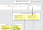
The bacteriological research method includes:
1. inoculation of the test material in nutrient media
2. pure culture isolation
3. identification of microorganisms (determination of belonging to a species).
Isolation and identification of pure cultures of aerobic and anaerobic bacteria involves the following studies:
Stage I (work with native material)
Purpose: obtaining isolated colonies
1. Preliminary microscopy gives an approximate idea of the microflora
2. Preparation of material for research
3. Seeding on dense nutrient media to obtain isolated colonies
4. Incubation at optimal temperature, most often 37°C, for 18-24 hours
II stage
Purpose: obtaining a pure culture
1. Macroscopic study of colonies in transmitted and reflected light (characterization of the size, shape, color, transparency, consistency, structure, contour, surface of the colonies).
2. Microscopic examination of isolated colonies
3. Testing for aerotolerance (to confirm the presence of strict anaerobes in the test material).
4. Sowing colonies characteristic of a certain species on pure culture accumulation media or elective media and incubation under optimal conditions.
Stage III
Purpose: Identification of isolated pure culture
1. To identify the isolated culture by a complex of biological properties, the following is studied:
morphology and tinctorial properties
cultural properties (nature of growth on nutrient media)
biochemical properties (enzymatic activity of microorganisms)
serological properties (antigenic)
virulent properties (ability to produce pathogenicity factors: toxins, enzymes, defense and aggression factors)
pathogenicity for animals
phage lizability (sensitivity to diagnostic bacteriophages)
sensitivity to antibiotics
other individual properties
Stage IV (Conclusion)
According to the studied properties, a conclusion is made about the isolated culture
The first stage of research. The study of pathological material begins with microscopy. Microscopy of stained native material makes it possible to establish approximately the composition of the microbial landscape of the studied object, some morphological features of microorganisms. The results of microscopy of native material largely determine the course of further research, and subsequently they are compared with the data obtained during inoculation on nutrient media.
With a sufficient content of pathogenic microorganisms in the sample, inoculation is carried out on dense nutrient media (to obtain isolated colonies). If there are few bacteria in the test material, then the inoculation is carried out on liquid nutrient enrichment media. Nutrient media are selected according to the requirements of microorganisms.
Cultivation of microorganisms is possible only when creating optimal conditions for their vital activity and observing the rules that exclude contamination (accidental contamination by foreign microbes) of the test material. Artificial conditions that would exclude culture contamination by other species can be created in a test tube, flask or Petri dish. All utensils and nutrient media must be sterile and, after inoculation of microbial material, protected from outside contamination, which is achieved using stoppers or metal caps and lids. Manipulations with the test material should be carried out in the flame zone of an alcohol lamp to exclude contamination of the material from the external environment, as well as in order to comply with safety regulations.
Inoculation of the material on nutrient media should be done no later than 2 hours from the moment of their collection.
The second stage of research. Study of colonies and isolation of pure cultures. After a day of incubation, colonies grow on the plates, and on the first stroke the growth is continuous, and on the next - isolated colonies. A colony is a collection of microbes of the same species that have grown from a single cell. Since the material is most often a mixture of microbes, several types of colonies grow. Different colonies are marked with a pencil, outlining them with a circle from the side of the bottom, and studying them (Table 11). First of all, study the colonies with the naked eye: macroscopic signs. The cup is viewed (without opening it) from the bottom in transmitted light, the transparency of the colonies is noted (transparent if it does not trap light; translucent if it partially traps light; opaque if light does not pass through the colony), measure (in mm) the size of the colonies. Then they study the colonies from the side of the lid, note the shape (regular round, irregular, flat, convex), the nature of the surface (smooth, shiny, dull, rough, wrinkled, wet, dry, mucous), color (colorless, colored).
Table 11. Scheme of the study of colonies
|
Possible colony characteristics |
||
|
Flat, convex, dome-shaped, depressed, round, rosette-shaped, star-shaped |
||
|
Size, mm |
Large (4-5 mm), medium (2-4 mm), small (1-2 mm), dwarf (< 1 мм) |
|
|
Surface nature |
Smooth (S-shape), rough (R-shape), slimy (M-shape), striated, bumpy, matte, shiny |
|
|
Colorless, dyed |
||
|
Transparency |
Transparent, opaque, translucent |
|
|
The nature of the edges |
Smooth, serrated, fringed, fibrous, scalloped |
|
|
Internal structure |
Homogeneous, granular, heterogeneous |
|
|
Consistency |
Viscous, slimy, crumbly |
|
|
Emulsification in a drop of water |
Good bad |
Note: 5-7 points are studied at low magnification of the microscope.
You can see the difference between the colonies even better when you zoom in on them. To do this, a closed dish is placed upside down on an object table, the condenser is slightly lowered, a small objective magnification (x8) is used, moving the dish, microscopic signs are studied in the colonies: the nature of the edge (smooth, wavy, serrated, scalloped), structure (homogeneous, granular, fibrous, homogeneous, or different in the center and along the periphery).
Next, the morphology of microbial cells from the colonies is studied. To do this, smears are made from a part of each of the marked colonies, stained according to Gram. When taking colonies, pay attention to the consistency (dry, if the colony crumbles and is taken with difficulty; soft, if it is taken easily on the loop; slimy, if the colony reaches for the loop; hard, if part of the colony is not taken by the loop, only the entire colony can be removed) .
When viewing smears, it is established that the colony is represented by one type of microbe, therefore, pure cultures of bacteria can be isolated. To do this, from the studied colonies, reseeding is done on a slant agar. When reseeding from colonies, care must be taken to take exactly the intended colonies, without touching the loop of nearby colonies. The tubes are signed and incubated in a thermostat at 37°C for 24 hours.
The third stage of research. Identification of the isolated culture. Identification of microbes - determination of the systematic position of the culture isolated from the material to the species and variant. The first condition for the reliability of identification is the unconditional purity of the culture. To identify microbes, a set of signs is used: morphological (shape, size, presence of flagella, capsules, spores, relative position in a smear), tinctorial (relationship to Gram stain or other methods), chemical (guanine + cytosine ratio in DNA molecule), cultural (nutrient requirements, cultivation conditions, growth rate and nature on various nutrient media), enzymatic (splitting various substances with the formation of intermediate and final products), serological (antigenic structure, specificity), biological (virulence for animals, toxigenicity, allergenicity, the effect of antibiotics, etc.).
For biochemical differentiation, the ability of bacteria to ferment carbohydrates with the formation of intermediate and end products, the ability to degrade proteins and peptones, and study redox enzymes.
To study saccharolytic enzymes, isolated cultures are inoculated into test tubes with semi-liquid media containing lactose, glucose and other carbohydrates and polyhydric alcohols. On semi-liquid media, inoculation is done by injection into the depth of the medium. When sowing by injection, the test tube with the medium is held at an angle, the stopper is removed, and the edge of the test tube is burned. The material is taken with a sterile loop and a column of nutrient medium is pierced almost to the bottom with it.
To determine proteolytic enzymes, the isolated culture is inoculated on peptone water or MPB. To do this, they take a test tube with inoculation closer to themselves, and a test tube with the medium - further away from themselves. Both test tubes are opened at the same time, capturing their stoppers with the little finger and the edge of the palm, the edges of the tubes are burned, a little culture is captured with a calcined cooled loop and transferred to the second test tube, triturated in a liquid medium on the wall of the test tube and washed off with the medium.
When sowing and reseeding, attention should be paid to compliance with the rules of sterility, in order not to contaminate your crops with foreign microflora, and also not to contaminate environment. The tubes are labeled and placed in a thermostat for incubation at 37°C for a day.
Conclusion
Accounting for results. Research conclusion. The results of identification are taken into account and, based on the totality of the data obtained, based on the classification and characteristics of the type strains described in the manual (Bergy's guide, 1994-1996), the type of isolated cultures is determined.
Bacteriological research method. Isolation of a pure culture of aerobic bacteria (1-2 stages)
1. The essence of the bacteriological method
3. Methods for obtaining isolated colonies
4. Methods for obtaining pure culture
1. Carry out primary seeding by the Koch and Drygalski method
2. Isolate pure culture
Work plan:
1. Bacteriological research method
2. Stages of the bacteriological method
3. Concept: pure culture, strain, colony
4. Methods for obtaining isolated colonies
5. Methods for obtaining a pure culture of aerobic bacteria
The main method of laboratory diagnosis of infectious diseases is the bacteriological method. The essence of the bacteriological method is the isolation of pathogens from the material under study and its identification (definition of the genus, species, varieties).
The bacteriological method allows you to determine:
1. Type of pathogen and make a diagnosis of the disease
2. Antibiotics that kill the pathogen and prescribe the appropriate treatment
3. Phagovars and serovars of the pathogen to identify the source of infection
The first stage is the primary inoculation of the test material to obtain isolated colonies. Primary sowing is carried out on storage media and DDS (cup media).
The second stage is the characterization of isolated colonies and obtaining a pure culture from them by reseeding onto a slanted column of the main medium.
The third stage is the identification of a pure culture - the determination of the genus, species, varieties of the pathogen, the determination of antibiotic sensitivity, the determination of fagovars.
The culture of microorganisms is bacteria grown on nutrient media. A pure culture is one species of bacteria grown on nutrient media. Within a species, there are varieties that differ in one trait:
1. Morphovars - differ in morphology
2. Chemovars - differ in biochemical properties
3. Serovars - differ in antigenic properties
4. Fagovars - differ in sensitivity to phages
A pure culture is necessary for identification, determination of serovars, phage, sensitivity to antibiotics. A strain is one species of bacteria isolated from a specific source (Volga River, sick Ivanov, etc.). Colony - a visible accumulation of bacteria of the same species, formed during the reproduction of one bacterial cell on a dense nutrient medium.
The first stage of the study is the primary inoculation of the test material to obtain isolated colonies. Before the initial inoculation, microscopy of the test material is carried out. The test material usually contains a mixture of various bacteria, including pathogens. To isolate the pathogen, it is necessary to obtain isolated colonies (bacteria of the same species) by selecting a nutrient medium and using special seeding methods.
There are two methods for obtaining isolated colonies:
1. Drygalski method