Proteins play a very important role in the life of organisms, perform protective, structural, hormonal, energy functions. Provides growth of muscle and bone tissue. Proteins inform about the structure of the cell, about its functions and biochemical properties, are part of the valuable, beneficial to the body food (eggs, dairy products, fish, nuts, legumes, rye and wheat). The digestibility of such food is explained by the biological value. With an equal indicator of the amount of protein, it will be easier to digest the product whose value is higher. Defective polymers must be removed from the body and replaced with new ones. This process occurs during the synthesis of proteins in cells.
What are proteins
Substances that consist only of amino acid residues are called simple proteins (proteins). If necessary, their energy property is used, therefore, people leading healthy lifestyle life, often additional protein intake is needed. Complex proteins, proteids, are composed of a simple protein and a non-protein part. Ten amino acids in a protein are essential, which means that the body cannot synthesize them on its own, they come from food, while the other ten are non-essential, that is, they can be created from other amino acids. This is how the process, vital for all organisms, begins.

The main stages of biosynthesis: where do proteins come from
New molecules are taken as a result of biosynthesis - a chemical reaction of a compound. There are two main steps in the synthesis of proteins in a cell. This is transcription and translation. Transcription takes place in the nucleus. This is a reading from DNA (deoxyribonucleic acid), which carries information about the future protein, to RNA (ribonucleic acid), which transfers this information from DNA to the cytoplasm. This happens due to the fact that DNA does not directly participate in biosynthesis, it only carries information, not being able to enter the cytoplasm where the protein is synthesized, and performing only the function of a carrier of genetic information. Transcription, on the other hand, makes it possible to read data from a DNA template into RNA according to the principle of complementarity.

The role of RNA and DNA in the process
So, it starts the synthesis of proteins in cells by a DNA chain that carries information about a particular protein and is called a gene. The DNA chain unwinds during transcription, that is, its helix begins to disintegrate into a linear molecule. From DNA information must be converted to RNA. In this process, adenine should become opposite thymine. Cytosine has guanine as a pair, just like DNA. Opposite adenine, RNA becomes uracil, because in RNA such a nucleotide as thymine does not exist, it is replaced simply by a uracil nucleotide. Cytosine is adjacent to guanine. Opposite adenine is uracil, and paired with thymine is adenine. These RNA molecules that stand opposite are called messenger RNA (mRNA). They are able to exit the nucleus through the pores into the cytoplasm and ribosomes, which, in fact, perform the function of protein synthesis in cells.
About the complex in simple words
Now, the assembly of the amino acid sequences of the polypeptide chain of the protein is being made. Transcription can be called the reading of information about the future protein from the DNA template into RNA. This can be defined as the first stage. After the RNA leaves the nucleus, it must reach the ribosomes, where the second step, called translation, takes place.

Translation is already the transition of RNA, that is, the transfer of information from nucleotides to a protein molecule, when RNA tells what sequence of amino acids should be in the substance. In this order, messenger RNA enters the cytoplasm to ribosomes that synthesize proteins in the cell: A (adenine) - G (guanine) - U (uracil) - C (cytosine) - U (uracil) - A (adenine).
Why are ribosomes needed?
In order for translation to occur and result in a protein, components such as messenger RNA itself, transfer RNA, and ribosomes as a "factory" in which protein is produced are needed. In this case, two types of RNA function: informational, which was formed in the nucleus with DNA, and transport. The second acid molecule looks like a clover. This "clover" attaches an amino acid to itself and carries it to the ribosomes. That is, it carries out transportation organic compounds directly to the "factory" of their education.
How rRNA works
There are also ribosomal RNAs that are part of the ribosome itself and perform protein synthesis in the cell. It turns out that ribosomes are non-membrane structures, they do not have shells, such as the nucleus or endoplasmic reticulum, but simply consist of proteins and ribosomal RNA. What happens when a sequence of nucleotides, that is, messenger RNA, gets to the ribosomes?

Transfer RNA, which is located in the cytoplasm, pulls amino acids to itself. Where did the amino acids in the cell come from? And they are formed due to the breakdown of proteins that are ingested with food. These compounds are transported by the blood stream to the cells, where the proteins necessary for the body are produced.
The final step in protein synthesis in cells
Amino acids swim in the cytoplasm in the same way as transfer RNAs, and when the assembly of the polypeptide chain occurs directly, these transfer RNAs begin to connect with them. However, not in any sequence and not any transfer RNA can combine with all types of amino acids. There is a specific site to which the necessary amino acid is attached. The second segment of the transfer RNA is called the anticodon. This element consists of three nucleotides that are complementary to the nucleotide sequence in messenger RNA. One amino acid requires three nucleotides. For example, any conditional protein consists, for simplicity, of only two amino acids. Obviously, most proteins have a very long structure, consisting of many amino acids. The chain A - G - Y is called a triplet, or codon, it will be joined by transfer RNA in the form of a clover, at the end of which there will be a certain amino acid. The next C-U-A triplet will be joined by another tRNA, which will contain a completely different amino acid complementary to this sequence. In this order, further assembly of the polypeptide chain will occur.

The biological significance of synthesis
Between the two amino acids located at the ends of the "clovers" of each triplet, a peptide bond is formed. At this stage, the transfer RNA goes into the cytoplasm. Then the next transport RNA with another amino acid joins the triplets, which forms a polypeptide chain with the previous two. This process is repeated until the required amino acid sequence is reached. Thus, protein synthesis occurs in the cell, and enzymes, hormones, blood substances, etc. are formed. Not every cell produces any protein. Each cell can form a specific protein. For example, hemoglobin will be formed in erythrocytes, and hormones and various enzymes will be synthesized by pancreatic cells that break down food that enters the body.
The proteins actin and myosin will be formed in the muscles. As can be seen, the process of protein synthesis in cells is multi-stage and complex, which indicates its importance and necessity for all living things.
Protein biosynthesis in cells is a sequence of matrix-type reactions, during which the successive transfer of hereditary information from one type of molecule to another leads to the formation of polypeptides with a genetically determined structure.
Protein biosynthesis is the initial stage of realization or expression of genetic information. The main matrix processes that ensure protein biosynthesis are DNA transcription and mRNA translation. DNA transcription consists in rewriting information from DNA to mRNA (messenger or messenger RNA). Translation of mRNA is the transfer of information from mRNA to a polypeptide. The sequence of matrix reactions in protein biosynthesis can be represented as a diagram.
untranscribed strand of DNA
transcribed DNA strand
DNA transcription
mRNA codons
mRNA translation
tRNA anticodons
protein amino acids
methionine
The diagram shows that the genetic information about the structure of a protein is stored as a sequence of DNA triplets. In this case, only one of the DNA chains serves as a template for transcription (such a chain is called transcribed). The second strand is complementary to the transcribed strand and is not involved in mRNA synthesis.
The mRNA molecule serves as a template for polypeptide synthesis on ribosomes. The mRNA triplets that code for a specific amino acid are called codons. Translation is carried out by tRNA molecules. Each tRNA molecule contains an anticodon, a recognition triplet in which the nucleotide sequence is complementary to a specific mRNA codon. Each tRNA molecule is capable of carrying a strictly defined amino acid. The combination of tRNA with an amino acid is called aminoacyl-tRNA.
The tRNA molecule in general conformation resembles a clover leaf on a petiole. The "top of the leaf" carries the anticodon. There are 61 types of tRNA with different anticodons. An amino acid is attached to the "leaf petiole" (there are 20 amino acids involved in the synthesis of the polypeptide on ribosomes). Each tRNA molecule with a certain anticodon corresponds to a strictly defined amino acid. At the same time, a certain amino acid usually corresponds to several types of tRNA with different anticodons. The amino acid covalently attaches to tRNA with the help of enzymes - aminoacyl-tRNA synthetases. This reaction is called tRNA aminoacylation.
On ribosomes, the anticodon of the corresponding aminoacyl-tRNA molecule is attached to a specific mRNA codon with the help of a specific protein. This binding of mRNA and aminoacyl-tRNA is called codon-dependent. On ribosomes, amino acids are connected to each other using peptide bonds, and the released tRNA molecules go in search of free amino acids.
Let us consider in more detail the main stages of protein biosynthesis.
Stage 1. DNA transcription. On the transcribed DNA strand, a complementary mRNA strand is completed using DNA-dependent RNA polymerase. The mRNA molecule is an exact copy of the non-transcribed DNA chain, with the difference that instead of deoxyribonucleotides it contains ribonucleotides, which include uracil instead of thymine.
Stage 2. Processing (maturation) of mRNA. The synthesized mRNA molecule (primary transcript) undergoes additional transformations. In most cases, the original mRNA molecule is cut into separate fragments. Some fragments - introns - are cleaved to nucleotides, while others - exons - are fused into mature mRNA. The process of connecting exons "without knots" is called splicing.
Splicing is characteristic of eukaryotes and archaebacteria, but sometimes also occurs in prokaryotes. There are several types of splicing. The essence of alternative splicing is that the same regions of the original mRNA can be both introns and exons. Then one and the same DNA region corresponds to several types of mature mRNA and, accordingly, several different forms of the same protein. The essence of trans splicing is the joining of exons encoded by different genes (sometimes even from different chromosomes) into one mature mRNA molecule.
Stage 3. mRNA translation. Translation (like all matrix processes) includes three stages: initiation (beginning), elongation (continuation) and termination (end).
Initiation. The essence of initiation is the formation of a peptide bond between the first two amino acids of the polypeptide.
Initially, an initiating complex is formed, which includes: a small subunit of the ribosome, specific proteins (initiation factors) and a special initiating methionine tRNA with the amino acid methionine - Met-tRNAMet. The initiating complex recognizes the beginning of the mRNA, attaches to it, and slides to the point of initiation (beginning) of protein biosynthesis: in most cases, this is the start codon AUG. Between the start codon of mRNA and the anticodon of methionine tRNA, codon-dependent binding occurs with the formation of hydrogen bonds. Then the large subunit of the ribosome is attached.
When the subunits combine, a complete ribosome is formed, which carries two active centers (sites): A-site (aminoacyl, which serves to attach aminoacyl-tRNA) and P-site (peptidyl transferase, which serves to form a peptide bond between amino acids).
Initially, Met-tRNAMet is located in the A-site, but then moves to the P-site. The vacated A site receives aminoacyl-tRNA with an anticodon that is complementary to the mRNA codon following the AUG codon. In our example, this is Gly-tRNAGly with the anticodon CCG, which is complementary to the GHC codon. As a result of codon-dependent binding, hydrogen bonds are formed between the mRNA codon and the aminoacyl-tRNA anticodon. Thus, two amino acids are adjacent to the ribosome, between which a peptide bond is formed. The covalent bond between the first amino acid (methionine) and its tRNA is broken.
After the formation of a peptide bond between the first two amino acids, the ribosome shifts by one triplet. As a result, translocation (movement) of the initiating methionine tRNAMet occurs outside the ribosome. The hydrogen bond between the start codon and the anticodon of the initiator tRNA is broken. As a result, free tRNAMet is cleaved off and goes in search of its amino acid.
The second tRNA, together with the amino acid (in our example, Gly-tRNAGly), as a result of translocation, ends up in the P-site, and the A-site is released.
Elongation. The essence of elongation is the addition of subsequent amino acids, that is, the extension of the polypeptide chain. The work cycle of the ribosome during elongation consists of three steps: codon-dependent binding of mRNA and aminoacyl-tRNA at the A site, formation of a peptide bond between the amino acid and the growing polypeptide chain, and translocation with the release of the A site.
The vacant A-site receives aminoacyl-tRNA with an anticodon corresponding to the next mRNA codon (in our example, this is Tir-tRNATir with the AUA anticodon, which is complementary to the UAU codon).
On the ribosome, two amino acids are next to each other, between which a peptide bond is formed. The link between the previous amino acid and its tRNA (in our example, between glycine and tRNAGly) is broken.
Then the ribosome moves one more triplet, and as a result of translocation, the tRNA that was in the P-site (in our example, tRNAgli) is outside the ribosome and is cleaved off from the mRNA. The A site is released, and the ribosome cycle starts over.
Termination. It consists in the completion of the synthesis of the polypeptide chain.
Eventually, the ribosome reaches an mRNA codon that no tRNA (and no amino acid) matches. There are three such nonsense codons: UAA (“ocher”), UAG (“amber”), UGA (“opal”). At these mRNA codons, the working cycle of the ribosome is interrupted, and the growth of the polypeptide stops. The ribosome, under the influence of certain proteins, is again divided into subunits.
Protein modification. As a rule, the synthesized polypeptide undergoes further chemical transformations. The original molecule can be cut into separate fragments; then some fragments are crosslinked, others are hydrolyzed to amino acids. Simple proteins can combine with a wide variety of substances, forming glycoproteins, lipoproteins, metalloproteins, chromoproteins and other complex proteins. In addition, amino acids already in the composition of the polypeptide can undergo chemical transformations. For example, the amino acid proline, which is part of the procollagen protein, is oxidized to hydroxyproline. As a result, collagen is formed from procollagen - the main protein component of connective tissue.
Protein modification reactions are not matrix-type reactions. Such biochemical reactions are called stepwise.
Energy of protein biosynthesis. Protein biosynthesis is a very energy intensive process. Aminoacylation of tRNA consumes the energy of one bond ATP molecules, with codon-dependent binding of aminoacyl-tRNA - the energy of one bond of the GTP molecule, when the ribosome moves one triplet - the energy of one bond of another GTP molecule. As a result, about 90 kJ / mol is spent on attaching an amino acid to a polypeptide chain. Hydrolysis of the peptide bond releases only 2 kJ/mol. Thus, during biosynthesis, most of the energy is irretrievably lost (dissipated in the form of heat).
Genetic code, its main properties
During the reactions matrix synthesis on the basis of the genetic code, a polypeptide with a hereditarily determined structure is synthesized. A segment of DNA containing information about the structure of a particular polypeptide is called a gene.
However, gene - this is not just a section of DNA, but a unit of hereditary information, the carrier of which are nucleic acids. It has been established that the gene has a complex structure.
In most cases, coding regions (exons) are separated by non-coding regions (introns). At the same time, due to alternative splicing, the division of a DNA segment into coding and non-coding turns out to be conditional. Some sections of DNA can move relative to each other - they are called mobile genetic elements (MGE). Many genes are represented by several copies - then the same protein is encoded by different sections of DNA. The genetic information of viruses is even more difficult to encode. Many of them have overlapping genes: the same piece of DNA can be transcribed from different starting points.
The process of gene expression is flexible: several polypeptides can correspond to one piece of DNA; one polypeptide can be encoded by different regions of DNA. The final modification of proteins occurs with the help of enzymes that are encoded by different sections of DNA.
General properties of the genetic code
The reflection of some objects with the help of others is called encoding. The reflection of the structure of proteins in the form of DNA triplets is called the DNA code, or the genetic code. Thanks to the genetic code, a one-to-one correspondence between nucleotide sequences is established. nucleic acids and amino acids that make up proteins. The genetic code has the following main properties:
1. The genetic code is triplet: each amino acid is encoded by a triplet of DNA nucleotides and the corresponding triplet of mRNA. At the same time, codons are not separated from each other in any way (there are no "commas").
2. The genetic code is redundant (degenerate): almost all amino acids can be encoded by different codons. Only two amino acids correspond to one codon each: methionine (AUG) and tryptophan (UGG). But leucine, serine and arginine correspond to 6 different codons.
3. The genetic code is non-overlapping: each pair of nucleotides belongs to only one codon (exceptions are found in viruses).
4. The genetic code is the same for the vast majority of biological systems. However, there are exceptions, for example, in ciliates and in mitochondria of various organisms. Therefore, the genetic code is called quasi-universal.
The biosynthesis of proteins (polypeptides) is an extremely complex and amazing process. Protein biosynthesis actively proceeds in all organs and tissues, excluding erythrocytes. Many cells synthesize proteins for "export" (cells of the liver, pancreas), and in this case they contain very big number ribosome. In an animal cell, the number of ribosomes reaches 105, the diameter of the ribosome is 20 nm.
The process of protein synthesis occurs inside cells on the surface of ribosomes, which are complexes of two subunits with a sedimentation constant of 60S and 40S, functioning as a single whole. The ribosome contains 30-35% protein and 65-70% ribosomal RNA. The ribosome has aminoacyl and peptidyl regions. The first serves to fix the active amino acid and tRNA complex that enters the ribosome, and the second fixes the polypeptide chain associated with another tRNA. Ribosome subunits are synthesized in the nucleolus of the nucleus on a DNA template.
The essence of the process of protein synthesis is the scheme:
The protein-synthesizing system includes ribosomes, nucleic acids, a set of 20 amino acids, various enzymes, ATP, GTP, magnesium ions, and about 200 different non-catalytic protein factors.
A protein molecule is a long chain of amino acid residues, averaging 100 to 500 amino acids. The synthesis program for each protein is stored in a deoxyribonucleic acid (DNA) molecule. The DNA molecule is a polymer whose monomers are nucleotides. The sequence of nitrogenous bases in a DNA molecule determines the sequence of amino acids in a protein molecule.
There are four types of nitrogenous bases in a DNA molecule: adenine (A), guanine (G), cytosine (C) and thymine (T). A sequence of three bases (a triplet) makes up a codon, which corresponds to one specific amino acid.
Nucleic acids - DNA and RNA - are essential components of protein biosynthesis. DNA is responsible for storing genetic information, while RNA determines the transmission of this information and implementation in the form of protein molecules. It can be argued that the main function of DNA is the preservation of the genotype, and RNA is the expression of this genotype.
In quantitative terms, ribosomal RNA (rRNA) predominates in the cell. rRNA has helical sections and contains modified nucleotides (for example, 2-methylribose). rRNA makes up about 80% of total RNA in the cell. The second type of RNA in the cell is transfer RNA (tRNA), which, like all other types of RNA, is synthesized in the nucleus. It accounts for 10-15% of the total amount of RNA in the cell. Over 60 different tRNAs have been identified. Therefore, there are several different tRNAs for the transport of individual amino acids. For every amino acid in a cell, there is at least one specific tRNA. tRNA molecules are relatively small. Their structure contains 75-93 ribonucleotides.
The amino acid is attached to the free 3-OH group of the terminal tRNA mononucleotide, which is always represented by adenylic acid. tRNA also has another important site - an anticodon, with the help of which the complex of amino acids and tRNA recognizes a certain sequence of three nucleotides in messenger RNA (codon). The anticodon and codon are complementary connected by hydrogen bonds.
If the carrier of hereditary information in a cell is DNA, which is concentrated in the nucleus, but protein synthesis occurs in the cytoplasm, then, therefore, there must be a certain mediator that transmits this information to the cytoplasm of the cell. This mediator turned out to be messenger or messenger RNA (mRNA). mRNA accounts for 2% of the total amount of RNA in a cell. mRNA molecules are the longest (include up to 5 thousand nucleotides). mRNA also contains four types of nitrogenous bases. Of these, three (A, G, C) are the same as in DNA, and the fourth is uracil.
The information encoded in mRNA is necessary for the synthesis of a protein molecule, which occurs on ribosomes. The synthesis of mRNA in the cell nucleus is very fast, which is necessary for the active biosynthesis of protein molecules. mRNA is formed on one of the DNA strands of the nucleus. In this case, the double-stranded structure of DNA unwinds, and with the participation of DNA-dependent RNA polymerase, according to the principle of complementarity, mRNA synthesis occurs:

mRNA synthesis scheme
The principle of complementarity means that adenine on the DNA helix corresponds to uracil mRNA, thymine to adenine, and guanine to cytosine. Therefore, mRNA reads information from DNA.
The DNA -» RNA stage thus determines the synthesis of an mRNA molecule in which the nucleotide sequence is complementary to a specific DNA region (gene). This process is called transcription. The mRNA then enters the ribosome, combining with its subunits. One mRNA molecule is fixed on many ribosomes at the same time, forming the so-called polysomes. The presence of polysomes increases the efficiency and speed of mRNA utilization.
The synthesis of a polypeptide chain of a certain composition occurs on an mRNA template. The process of transferring information from mRNA to protein is called translation. The RNA -> protein step represents the process of protein synthesis directed by mRNA. Thus, the transfer of information always goes in the direction of DNA -» RNA -» protein.
The translation process includes the following steps:
- 1) activation of amino acids and their fixation on tRNA;
- 2) initiation of the synthesis of the polypeptide chain;
- 3) elongation of the synthesized polypeptide chain;
- 4) termination of the polypeptide chain and its release;
- 5) post-translational modification of the polypeptide chain.
- 1. Activation of amino acids requires the enzyme aminoacyl-tRNA synthetase and energy expenditure in the form of ATP:
The same enzyme is involved in fixing the previously activated amino acid to position 2 or 3 of the ribose of the last tRNA nucleotide:
In the form of this complex, the amino acid is transported to the ribosome, where the protein molecule is synthesized. Aminoacyl-tRNA synthetase is specific; it is able to recognize both amino acids and tRNAs. In a cell, therefore, there are at least 20 different synthetases, in accordance with the number of a-amino acids.
2. tRNA, linked by an ester bond with a certain amino acid, enters the ribosome and interacts with mRNA according to the type of complementarity between a specific triplet of mRNA nucleotides, called a codon, and its complementary specific triplet of nucleotides (anticodon) of a tRNA carrying a specific amino acid. Thus, each mRNA codon corresponds to a specific fixation of one amino acid in the peptide chain by the tRNA anticodon. The ribosome moves along the mRNA molecule, reading all the codons in sequence, thus establishing the order of all amino acids delivered to the site of synthesis.
The synthesis of a protein molecule proceeds in the direction from the free amino group to the free carboxyl group of the amino acid. Usually, the initial amino acid in the synthesis of a polypeptide chain is methionine, for which the nucleotide sequence of AUG mRNA serves as a codon.
The initiation of polypeptide synthesis begins when two tRNA anticodons are fixed at the corresponding mRNA codons. The process requires the presence of an energy source, which is GTP, as well as the participation of a number of protein initiation factors and peptidyl transferase.
With the participation of this enzyme, the rate of formation covalent bonds reaches 1200 amino acids/min/ribosome.

Polypeptide synthesis initiation scheme
3. After the formation of the dipeptide, the “unloaded” tRNA leaves the ribosome and is able to deliver new amino acid molecules, and the mRNA advances three nucleotides relative to the ribosome (polysome). As a result of movement (translocation), a free codon occupies a position for recognition of the next tRNA molecule. Therefore, in the elongation stage, a sequential addition of one amino acid to the polypeptide chain occurs in strict accordance with the order of the codons of the mRNA molecule.
An elongating polypeptide chain with one tRNA molecule is fixed to the large subunit of the ribosome. The attachment of each additional amino acid to the polypeptide chain occurs due to the relationship between the amino group of the amino acid in the complex with tRNA and the carboxyl group of the peptide.
4. Termination, or completion of the synthesis of a polypeptide molecule, involves certain “senseless” termination codons and protein termination factors. Three codons are known (UAG, UGA, UAA) that do not encode, do not bind any amino acid, since there are no tRNA anticodons in the cell that are complementary to them. Theoretically, only one "senseless" codon recognized by the polysome during its passage in the direction of 5-3 mRNA should stop the synthesis of a protein molecule.
The presence of a termination codon in any region of mRNA means the end of protein synthesis. As a result, the polysome disintegrates, the unused mRNA is hydrolyzed by polynucleotide phosphorylase, and the ribosome subunits are prepared to begin the synthesis of a new protein molecule.
mRNA can repeatedly participate in the process of protein biosynthesis. The duration of the functioning of the mRNA molecule is not the same in different organisms. It can vary from several minutes to several days.
5. Only the primary structure of a protein is encoded in DNA. Therefore, protein molecules synthesized on ribosomes do not yet have a completely completed state. They are primary polypeptides, which then undergo numerous modifications (association of monomers to form oligomers, addition of coenzymes, chemical transformations) that change the structure of proteins and, therefore, their activity.
The secondary and tertiary structures are not encoded, they are determined by the properties of the primary structure, which means that one or another form of a protein molecule depends on the sequence of amino acids and the possibilities of their interaction with each other. Structural modifications of synthesized proteins take place at the level of ribosomes or after completion of synthesis as a result of the addition of various functional groups.
The considered scheme of information transfer in the form
may change in individual cases. So, in viruses that do not contain DNA, information is embedded in RNA. When a virus enters a cell, this information is transmitted to the DNA of the cell, and the latter already synthesizes mRNA, on the matrix of which viral proteins are synthesized. Such a process is called reverse transcription, and the information transfer scheme in this case will be as follows:
As long as the sequence of DNA nucleotides and, consequently, mRNA is preserved, the nature of the newly synthesized protein remains unchanged.
The necessary genetic information for protein synthesis can be represented similarly to the human language, which consists of a sequence of letters that form words and sentences. In the genetic language, however, there are only four letters - four bases (adenine, guanine, uracil, cytosine).
The genetic code includes three-letter words. The four bases in this case (43) give 64 variants (words), which are more than enough to code for 20 amino acids. Thus, 64 codons make up the genetic code (Table 3).
Analysis of the genetic code shows that there are different numbers of codons for different amino acids. For example, methionine and tryptophan have only one codon, while arginine, leucine, and serine each have six codons. The presence of several codons for one amino acid reflects the "degeneracy" of the code. Consequently, the same amino acid can be encoded by several nucleotide triplets in its structure. At the same time, each triplet corresponds to a well-defined amino acid in the synthesized polypeptide chain.
Table 3
Genetic code
|
nucleotide |
Second nucleotide |
nucleotide |
|||
The genetic code is universal and the same across species different levels development (human, animals, plants, microorganisms). The universality of the code indicates that all living organisms in the past had a single ancestor.
Individual amino acids (hydroxyproline, oxylysin), for example, do not have a codon and are formed using chemical reactions after the synthesis of the polypeptide chain. This process is called post-translational modification and is very important for the proper functioning of each protein.
Meaningless codons (UAA, UAG, UGA) do not code for amino acids, but actually serve as a signal for the end of the synthesis of a protein molecule.
Thus, mRNA is a direct carrier of genetic information from the nucleus to the cytoplasmic ribosome. One ribosome occupies a region of about 80 nucleotides in length on mRNA and is capable of catalyzing approximately 100 peptide bonds per minute (Severin E. S. et al., 2011).
Synthesized protein molecules can undergo structural modifications at the level of ribosomes or after completion of synthesis as a result of the addition of various functional groups. In the cytoplasm, mRNA has a relatively short period of existence. Some mRNA is synthesized and stored in an inactive form, ready for rapid protein synthesis. Since mRNA information is associated with a linear sequence of nucleotides, the integrity of this sequence is extremely important. Any loss or change in the order of nucleotides can alter protein synthesis. To date, a number of inhibitors of DNA replication in the cells of the body (antibiotics, chemical poisons, antiviral drugs) have been installed. Damage to the sequence of purine or pyrimidine bases in a gene is called a mutation.
Substitution of only one nucleotide in a codon (mutation) leads to a change in the coding of one amino acid for another. For example, a mutation associated with the replacement of glutamic acid with valine in the hemoglobin molecule leads to the synthesis of hemoglobin, which causes sickle-cell anemia. Today, more than 200 mutations of the polypeptide chain of the human hemoglobin molecule are known. Often mutagens are substances (nitrosamines, for example) that change the structure of nitrogenous bases, which leads to a change in the nature of base complementarity. Ultraviolet irradiation causes condensation of thymine residues to form thymine dimers. Fortunately, animals are protected from the harmful effects of ultraviolet rays by the ozone layer of the atmosphere.
Many antibiotics used in veterinary practice inhibit bacterial protein synthesis (lincomycin, erythromycin, chloramphenicol) even at the translation stage. In this case, the microbial cell dies or stops its development. Antibiotics such as tetracyclines do not affect ribosomal synthesis in higher animal cells. Penicillins are not direct inhibitors of protein synthesis, however, their bacterial inhibition effects are associated with blocking the synthesis of cell wall hexapeptides. It should be noted that protein synthesis occurs not only on ribosomes, but also in mitochondria. Mitochondria have a complete and independent protein synthesis apparatus for their needs, although not all mitochondrial proteins are synthesized in these organelles. Mitochondrial RNA makes up only 3% of the total cellular RNA. Mitochondrial ribosomes are smaller than cytoplasmic ones. The UGA codon, as a terminator of protein synthesis in the cytoplasm, is used in mitochondria along with the UGG codon to encode an amino acid.
Proteins synthesized on ribosomes do not yet have a completely completed state. They represent primary polypeptides, which then undergo numerous modifications (association of monomers to form oligomers, addition of coenzymes, chemical transformations) that modify the structure of the protein and, hence, its activity.
Introduction
Life is a way of existence of protein bodies. This definition, given by Friedrich Engels, indicates the exceptional role of proteins in the functioning of organisms. Protein biosynthesis is an extremely complex and energy-intensive process. It is the basis of cell life.
Protein synthesis is carried out in ribosomes and takes place in several stages according to the scheme DNARNA protein. A double-stranded DNA molecule is transcribed into a single-stranded RNA molecule based on the principle of complementarity. The result is messenger RNA, which contains information about the amino acid sequence of the protein. Then the mRNA enters the ribosome and, like a template, a protein is synthesized by it, by translating genetic information from the language of the nucleotide sequence into the language of the amino acid sequence. Step by step, a polypeptide chain is built, which, during and after synthesis, is modified into a biologically active protein. The synthesized protein is transported to different parts of the cell to perform its functions.
The coding of the amino acid sequence of proteins is carried out according to certain rules, called genetic code. Deciphering the genetic code is a very significant achievement of science. The code explains the mechanism of protein synthesis, the origin of mutations, and other biological phenomena.
X-ray diffraction analysis and others modern methods research has made it possible to advance far in the study of protein biosynthesis and other aspects molecular biology. Nevertheless, the spatial structures of some vitally important macromolecules have not yet been established. Science has to answer many questions regarding protein synthesis.
General scheme protein biosynthesis
General scheme of protein biosynthesis in a cell: DNARNA protein (Figure 1).
Figure 1. General scheme of protein biosynthesis in a cell
Transcription. Separate sections of double-stranded DNA (genes) serve as templates for the synthesis of single-stranded RNA chains on them according to the principle of complementarity. Transcription occurs in three stages: initiation, elongation, and termination.
processing and transport. In the process of synthesis, RNA undergoes changes, as a result of which it turns into a mature molecule suitable for protein synthesis. The resulting messenger RNA (mRNA) then enters the ribosomes as a program that determines the amino acid sequence in the synthesized protein.
Activation and acceptance of amino acids. Proteins are made up of amino acids, but free cell amino acids cannot be used directly by the ribosome. Each amino acid is first activated by ATP, and then attached to a special RNA molecule - transfer (transport) RNA (tRNA) outside the ribosome. The resulting aminoacyl-tRNA enters the ribosome as a substrate for protein synthesis.
Broadcast. The flow of information in the form of mRNA and the flow of material in the form of aminoacyl-tRNA enter the ribosomes, which translate (translate) genetic information from the language of the mRNA nucleotide sequence into the language of the amino acid. Each ribosome moves along the mRNA from one end to the other and, accordingly, selects from the environment those aminoacyl-tRNAs that correspond (complementary) to the triplet combinations of nucleotides that are currently in the ribosome. The amino acid residue of the selected aminoacyl-tRNA is each time covalently attached by the ribosome to the growing polypeptide chain, and the deacylated tRNA is released from the ribosome into solution. This is how the polypeptide chain is built sequentially.
Formation of a functional protein. During synthesis, the polypeptide chain is released from the ribosome and folded into a globule. Protein folding and transport are accompanied by enzymatic modifications (protein processing).
Despite the great complexity of the protein biosynthesis apparatus, it proceeds at an extremely high rate. The synthesis of thousands of different proteins in each cell is strictly ordered - under given metabolic conditions, only the necessary number of molecules of each protein is synthesized.
The most important functions of the body - metabolism, growth, development, transmission of heredity, movement, etc. - are carried out as a result of many chemical reactions involving proteins, nucleic acids and other biologically active substances. At the same time, various compounds are continuously synthesized in cells: building proteins, enzyme proteins, hormones. In the course of exchange, these substances wear out and are destroyed, and new ones are formed in their place. Since proteins create the material basis of life and accelerate all metabolic reactions, the vital activity of the cell and the organism as a whole is determined by the ability of cells to synthesize specific proteins. Their primary structure is predetermined by the genetic code in the DNA molecule.
Protein molecules consist of tens and hundreds of amino acids (more precisely, of amino acid residues). For example, there are about 600 of them in a hemoglobin molecule, and they are distributed into four polypeptide chains; in the ribonuclease molecule there are 124 such amino acids, etc.
Molecules play the main role in determining the primary structure of a protein DNA. Its different sections encode the synthesis of different proteins, therefore, one DNA molecule is involved in the synthesis of many individual proteins. The properties of proteins depend on the sequence of amino acids in the polypeptide chain. In turn, the alternation of amino acids is determined by the sequence of nucleotides in DNA, and each amino acid corresponds to a certain triplet. It has been experimentally proven that, for example, a DNA region with an AAC triplet corresponds to the amino acid leucine, an ACC triplet to tryptophan, an ACA triplet to cysteine, and so on. By dividing the DNA molecule into triplets, one can imagine which amino acids and in what sequence will be located in the protein molecule. The set of triplets makes up the material basis of genes, and each gene contains information about the structure of a specific protein (a gene is the basic biological unit of heredity; in chemical terms, a gene is a DNA segment that includes several hundred base pairs).
genetic code - the historically established organization of DNA and RNA molecules, in which the sequence of nucleotides in them carries information about the sequence of amino acids in protein molecules. Code properties: triplet (codon), non-overlapping (codons follow each other), specificity (one codon can determine only one amino acid in the polypeptide chain), universality (in all living organisms, the same codon determines the inclusion of the same amino acid in the polypeptide), redundancy (for most amino acids there are several codons). Triplets that do not carry information about amino acids are stop triplets, indicating the start of synthesis i-RNA.(V.B. Zakharov. Biology. Reference materials. M., 1997)
Since DNA is located in the nucleus of the cell, and protein synthesis occurs in the cytoplasm, there is an intermediary that transmits information from DNA to ribosomes. RNA also serves as such an intermediary, to which the nucleotide sequence is rewritten, in exact accordance with that on DNA - according to the principle of complementarity. This process has been named transcriptions and proceeds as a matrix synthesis reaction. It is characteristic only for living structures and underlies the most important property of living things - self-reproduction. Protein biosynthesis is preceded by template synthesis of mRNA on DNA strands. The resulting mRNA exits the cell nucleus into the cytoplasm, where ribosomes are strung on it, and amino acids are delivered here with the help of TRIC.
Protein synthesis is a complex multi-stage process involving DNA, mRNA, tRNA, ribosomes, ATP, and various enzymes. First, amino acids in the cytoplasm are activated by enzymes and attached to tRNA (to the site where the CCA nucleotide is located). The next step is the combination of amino acids in the order in which the alternation of nucleotides from DNA is transferred to mRNA. This stage is called broadcast. Not one ribosome is placed on the mRNA strand, but a group of them - such a complex is called a polysome (N.E. Kovalev, L.D. Shevchuk, O.I. Shchurenko. Biology for preparatory departments of medical institutes).
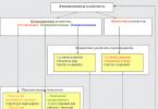
Scheme Protein biosynthesis
Protein synthesis consists of two stages - transcription and translation.
I. Transcription (rewriting) - biosynthesis of RNA molecules, carried out in chromosomes on DNA molecules according to the principle of matrix synthesis. With the help of enzymes, all types of RNA (mRNA, rRNA, tRNA) are synthesized at the corresponding sections of the DNA molecule (genes). 20 varieties of tRNA are synthesized, since 20 amino acids take part in protein biosynthesis. Then mRNA and tRNA exit into the cytoplasm, rRNA is integrated into ribosome subunits, which also exit into the cytoplasm.
II. Translation (transmission) - the synthesis of polypeptide chains of proteins, is carried out in ribosomes. It is accompanied by the following events:
1. Formation of the functional center of the ribosome - FCR, consisting of mRNA and two subunits of ribosomes. There are always two triplets (six nucleotides) of mRNA in PCR, which form two active centers: A (amino acid) - the amino acid recognition center and P (peptide) - the center for attaching the amino acid to the peptide chain.
2. Transportation of amino acids attached to tRNA from the cytoplasm to PCR. In the active center A, the tRNA anticodon is read with the mRNA codon; in the case of complementarity, a bond occurs that serves as a signal to advance (jump) along the mRNA of the ribosome by one triplet. As a result, the complex "codon of rRNA and tRNA with amino acid" moves to the active center of P, where the amino acid is attached to the peptide chain (protein molecule). The tRNA then leaves the ribosome.
3. The peptide chain lengthens until translation ends and the ribosome jumps off the mRNA. Several ribosomes (polysome) can fit on one mRNA at the same time. The polypeptide chain is immersed in the channel of the endoplasmic reticulum and there it acquires a secondary, tertiary or quaternary structure. The assembly speed of one protein molecule, consisting of 200-300 amino acids, is 1-2 minutes. Protein biosynthesis formula: DNA (transcription) --> RNA (translation) --> protein.
After completing one cycle, polysomes can take part in the synthesis of new protein molecules.
The protein molecule separated from the ribosome has the form of a thread that is biologically inactive. It becomes biologically functional after the molecule acquires a secondary, tertiary, and quaternary structure, i.e., a certain spatially specific configuration. The secondary and subsequent structures of a protein molecule are predetermined in the information embedded in the alternation of amino acids, i.e., in the primary structure of the protein. In other words, the program for the formation of a globule, its unique configuration, is determined by the primary structure of the molecule, which, in turn, is built under the control of the corresponding gene.
The rate of protein synthesis is determined by many factors: the temperature of the environment, the concentration of hydrogen ions, the amount final product synthesis, the presence of free amino acids, magnesium ions, the state of ribosomes, etc.