The repair mechanism is based on the presence of two complementary chains in the DNA molecule. Distortion of the nucleotide sequence in one of them is detected by specific enzymes. Then the corresponding section is removed and replaced with a new one, synthesized on the second complementary DNA strand. This type of repair is called excision repair, i.e. with “cutting” (Fig. 15). It occurs before the next replication cycle, which is why it is also called pre-replicative.
Fig. 14. Scheme of the correction process during DNA synthesis:
I-inclusion in the DNA chain of a nucleotide with an altered (tautomeric) form of cytoeine, which “illegally” pairs with adenine; II - the rapid transition of cytosine to its normal form disrupts its pairing with adenine; the unpaired 3"-OH end of the synthesized chain prevents its further elongation under the action of DNA polymerase; III - DNA polymerase removes the illegal nucleotide, as a result of which the 3"-OH end paired with the matrix reappears; IV - DNA polymerase continues to extend the chain at the 3"-OH end.
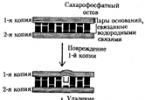
Restoring the original DNA structure requires the participation of a number of enzymes. An important point in triggering the repair mechanism is the detection of an error in the DNA structure. Often such errors occur in the newly synthesized chain during the replication process. Repair enzymes must detect this particular chain. In many species of living organisms, the newly synthesized DNA strand differs from the maternal one in the degree of methylation of its nitrogenous bases, which lags behind synthesis. In this case, the unmethylated chain undergoes repair. DNA strand breaks can also be recognized by repair enzymes. In higher organisms, where DNA synthesis does not occur continuously, but in separate replicons, the newly synthesized DNA strand has breaks, which makes it possible to recognize it. Restoring the DNA structure when the purine bases of one of its chains are lost involves detecting the defect using the enzyme endonuclease, which breaks the phosphoester bond at the site of damage to the chain. Then the changed section with several adjacent nucleotides is removed by the enzyme exonuclease, and in its place, in accordance with the order of the bases of the complementary chain, the correct nucleotide sequence is formed (Fig. 15).

Fig. 15. Scheme of excision, pre-replicative DNA repair.
When one of the bases in the DNA chain changes, about 20 DNA glycosylase enzymes take part in restoring the original structure. They specifically recognize damage caused by deamination, alkylation and other structural transformations of bases. Such modified bases are removed. Areas devoid of bases appear and are repaired, as with the loss of purines. If the normal structure is not restored, for example in the case of deamination of nitrogenous bases, some pairs of complementary bases are replaced by others - the C-G pair can be replaced by a T-A pair, etc. .
The formation of thymine dimers (T-T) in polynucleotide chains under the influence of UV rays requires the participation of enzymes that recognize not individual altered bases, but more extensive damage to the DNA structure. The repair process in this case is also associated with the removal of the region bearing the dimer and the restoration of the normal nucleotide sequence by synthesis on the complementary DNA strand.
In the case when the excision repair system does not correct a change that has arisen in one DNA strand, during replication this change is fixed and it becomes the property of both DNA strands. This leads to the replacement of one pair of complementary nucleotides with another or to the appearance of breaks (gaps) in the newly synthesized chain against the changed sections. Restoration of the normal DNA structure can also occur after replication.
DNA is a linear polymer containing from 70-80 to 10 9 mononucleotides, which are connected by covalent phosphodiester bonds that occur between the pentose hydroxyl group of one nucleotide and the phosphate group of the next nucleotide.
X-ray diffraction analysis data showed that the DNA molecule of most living organisms, with the exception of some phages, consists of two polynucleotide chains, antiparallel directed and oriented in such a way that their sugar-phosphate backbones are on the outside, and their nitrogenous bases are on the inside. The bases are arranged in pairs opposite each other and are connected by hydrogen bonds. Pairing occurs only between complementary (suitable to each other) bases: one purine and one perimedine. The A-T pair is connected by two, and the G-C pair by three hydrogen bonds. The DNA molecule has the shape of a double helix, in which the polynucleotide chains are twisted around an imaginary central axis.
The DNA helix is characterized by a number of parameters:
helix width is about 2 nm;
the pitch or complete turn of the helix is 3.4 nm and contains 10 pairs of complementary nucleotides.
DNA has unique properties: the ability to self-duplicate (replication) and the ability to self-heal (repair).
20 proteins: recognizing altered sections of DNA and removing them from the chain, restoring the correct sequence of nucleotides and stitching the restored fragment with the rest of the DNA molecule. 5% of all cellular RNA.
Replication carried out under the control of a number of enzymes and occurs in several stages. It begins at certain points in the DNA molecule. Special enzymes break the hydrogen bonds between complementary nitrogen bonds, and the helix unwinds. The polynucleotide chains of the parent molecule are kept in an untwisted state and serve as a template for the synthesis of new chains.
With the help of the enzyme DNA polymerase, daughter chains are assembled from the deoxyribonucleotide triphosphates (dATP, dGTP, dCTP, dTTP) available in the medium, complementary to the mother chains. Replication occurs simultaneously on both maternal strands, but at different speeds and with some differences. On one of the chains (leading) the assembly of the daughter chain occurs continuously, on the other (lagging) - fragmentarily. Subsequently, the synthesized fragments are mixed using the enzyme DNA ligase. As a result, two DNA molecules are formed from one DNA molecule, each of which has a mother and daughter chains. The synthesized molecules are exact copies of each other and the original DNA molecule. This method of DNA replication is called semi-conservative and ensures accurate reproduction in daughter molecules of the information that was recorded in the mother molecule.
Reparations is the ability of a DNA molecule to “correct” changes that occur in its chains. The restoration of the original DNA molecule involves at least
The listed features of the chemical structure and properties of DNA determine the functions it performs. DNA records, stores, reproduces genetic information, and participates in the processes of its implementation between new generations of cells and organisms.
Ribosomal RNA (rRNA) It is synthesized mainly in the nucleolus, in the region of rRNA genes, and is represented by molecules of various molecular weights that are part of the large or small subunits of ribosomes. rRNA accounts for 85% of the total RNA in a cell.
Transfer RNA (tRNA) makes up about 10% of cellular RNA. There are more than 40 types of tRNA. When implementing genetic information, each tRNA attaches a specific amino acid and transports it to the site of polypeptide assembly. In eukaryotes, tRNAs consist of 70-90 nucleotides and have a cloverleaf-shaped structure.
Ribonucleic acids - RNA - are represented by molecules of various sizes, structures and functions. All RNA molecules are copies of certain sections of the DNA molecule and, in addition to the differences already mentioned, are shorter than it and consist of a single chain. Between individual sections of one RNA chain that are complementary to each other, base pairing (A-U, G-C) and the formation of helical sections are possible. As a result, the molecules acquire a specific conformation.
Matrix, or information, RNA(mRNA, mRNA) synthesized in the nucleus under the control of the enzyme RNA polymerase, complementary to informative DNA sequences, transfers this information to ribosomes, where it becomes a matrix for the synthesis of a protein molecule. Depending on the amount of information copied, the mRNA molecule can have different lengths and makes up about 5% of all cellular RNA.
Table of contents of the topic "Genetic elements of bacteria. Mutations in bacteria. Transduction.":1. Migrating genetic elements of bacteria. Transposons. Bacteriophages as migrating genetic elements.
2. Mutation. Mutations in bacteria. Mutagens. Spontaneous mutations. Back mutations (reversions).
3. Induced mutations of bacteria. Chemical mutagenesis. Radiation mutagenesis. Types of mutations.
4. Bacterial DNA repair. DNA repair systems. Compensation for functions impaired as a result of mutations. Intragenic suppression. Extragenic suppression.
5. Transfer of bacterial DNA. Conjugation of bacteria. F-factor of bacteria.
6. Transformation of bacteria. Stages of bacterial transformation. Mapping bacterial chromosomes.
7. Transduction. Nonspecific transduction. Specific transduction. Abortive transduction. The phenomenon of lysogeny.
8. Properties of bacteria. Non-heritable changes in the properties of bacteria. S - colonies. R - colonies. M - colonies. D - colonies of bacteria.
Bacterial DNA repair. DNA repair systems. Compensation for functions impaired as a result of mutations. Intragenic suppression. Extragenic suppression.
There are mechanisms in the cell that can fully or partially restore the original structure of altered DNA. Mutations caused by radiation, chemicals and other factors could theoretically lead to the extinction of a bacterial population if the latter were deprived of the ability to DNA repair. A set of enzymes that catalyze the correction of DNA damage are combined into so-called repair systems, which differ fundamentally in the biochemical mechanisms of “healing” damage. There are three main directions for correcting DNA defects.
1. Direct reversal from damaged DNA to the original structure when changes in DNA corrected by a single enzymatic reaction. For example, removal of an incorrectly attached methyl group at the sixth oxygen atom of guanine using methyltransferase; or cleavage of the thymine dimer resulting from irradiation using photolyase ( recombinational repair).
2. "Cutting out" damage followed by restoration of the original structure (excision repair).
3. Activation of special mechanisms, ensuring survival in case of damage DNA(restoration of the original structure DNA as a result of recombination; correction of erroneous base pairing; translational synthesis on a damaged matrix DNA). These mechanisms do not always lead to complete restoration of the original DNA structure.
Compensation for functions impaired as a result of mutations
Primary mutation can be compensated by a secondary mutation that occurs within the mutated gene (intragenically) or in another gene (extragenically). Changes that eliminate the manifestations of a mutation without correcting the original disorder in DNA, called suppression.
Intragenic suppression caused by a secondary mutation correcting the effects of the primary mutation. For example, a point mutation that results in the synthesis of a defective protein with lost biological activity can be corrected if a secondary point mutation results in encoding an amino acid that retains the protein's configuration and activity. The exact restoration of the original gene structure is called a true reverse mutation ( true reversion). If the effect of the first mutation is compensated by a mutation in another part of the gene, such mutations are called secondary reversions.
Extragenic suppression- suppression of the manifestation of a mutation that occurred in one gene due to a mutation in the second gene.
Rice. 3.14. Scheme of the correction process during DNA synthesis:
I-inclusion in the DNA chain of a nucleotide with an altered (tautomeric) form of cytoeine, which “illegally” pairs with adenine; II - the rapid transition of cytosine to its normal form disrupts its pairing with adenine; the unpaired 3"-OH end of the synthesized chain prevents its further elongation under the action of DNA polymerase; III - DNA polymerase removes the illegal nucleotide, as a result of which the 3"-OH end paired with the matrix reappears; IV - DNA polymerase continues chain extension at the 3"-OH end
Restoring the original DNA structure requires the participation of a number of enzymes. An important point in triggering the repair mechanism is the detection of an error in the DNA structure. Often such errors occur in the newly synthesized chain during the replication process. Repair enzymes must detect this particular chain. In many species of living organisms, the newly synthesized DNA strand differs from the maternal one in the degree of methylation of its nitrogenous bases, which lags behind synthesis. In this case, the unmethylated chain undergoes repair. DNA strand breaks can also be recognized by repair enzymes. In higher organisms, where DNA synthesis does not occur continuously, but in separate replicons, the newly synthesized DNA strand has breaks, which makes it possible to recognize it.
Restoring the DNA structure when the purine bases of one of its chains are lost involves detecting the defect using the enzyme endonuclease, which breaks the phosphoester bond at the site of damage to the chain. Then the changed section with several adjacent nucleotides is removed by the enzyme exonuclease, and in its place, in accordance with the order of the bases of the complementary chain, the correct nucleotide sequence is formed (Fig. 3.15).
Rice. 3.15. Scheme of excision, pre-replicative DNA repair
When one of the bases in the DNA chain changes, about 20 DNA glycosylase enzymes take part in restoring the original structure. They specifically recognize damage caused by deamination, alkylation and other structural transformations of bases. Such modified bases are removed. Areas devoid of bases appear and are repaired, as with the loss of purines. If the normal structure is not restored, for example in the case of deamination of nitrogenous bases, some pairs of complementary bases are replaced by others - the C-G pair can be replaced by a T-A pair, etc. (see section 3.4.2.3).
The formation of thymine dimers (T-T) in polynucleotide chains under the influence of UV rays requires the participation of enzymes that recognize not individual altered bases, but more extensive damage to the DNA structure. The repair process in this case is also associated with the removal of the region carrying the dimer and the restoration of the normal nucleotide sequence by synthesis on the complementary DNA strand.
In the case when the excision repair system does not correct a change that has arisen in one DNA strand, during replication this change is fixed and it becomes the property of both DNA strands. This leads to the replacement of one pair of complementary nucleotides with another or to the appearance of breaks (gaps) in the newly synthesized chain against the changed sections. Restoration of the normal DNA structure can also occur after replication.
Post-replicative repair is carried out by recombination (exchange of fragments) between two newly formed DNA double helices. An example of such post-replicative repair is the restoration of normal DNA structure when thymine dimers (T-T) arise, when they are not spontaneously eliminated under the influence of visible light (light repair) or during pre-replicative excision repair.
Covalent bonds that arise between adjacent thymine residues make them incapable of binding to complementary nucleotides. As a result, breaks (gaps) appear in the newly synthesized DNA chain, recognized by repair enzymes. Restoration of the integrity of the new polynucleotide chain of one of the daughter DNA is carried out due to recombination with the corresponding normal parent chain of another daughter DNA. The gap formed in the mother chain is then filled by synthesis on a polynucleotide chain complementary to it (Fig. 3.16). A manifestation of such post-replicative repair, carried out by recombination between the chains of two daughter DNA molecules, can be considered the often observed exchange of material between sister chromatids (Fig. 3.17).
Rice. 3.16. Scheme of post-replicative DNA repair:
I - the appearance of a thymine dimer in one of the DNA chains;
II - formation of a “gap” in the newly synthesized chain against the changed section of the mother molecule after replication (the arrow shows the subsequent filling of the “gap” with a section from the corresponding chain of the second daughter DNA molecule);
III - restoration of the integrity of the daughter chain of the upper molecule due to recombination and in the lower molecule due to synthesis on the complementary chain
Rice. 3.17. Interchromatid exchanges (indicated by arrows)
During pre-replicative and post-replicative repair, most of the damage to the DNA structure is restored. However, if too much damage occurs in the hereditary material of the cell and some of it is not eliminated, the system of inducible (stimulated) repair enzymes (SOS system) is activated. These enzymes fill the gaps, restoring the integrity of the synthesized polynucleotide chains without strictly observing the principle of complementarity. This is why sometimes the repair processes themselves can serve as a source of permanent changes in the DNA structure (mutations). This reaction also applies to the SOS system.