Accent placement: ACTINOMYCETES
ACTINOMYCETES (Actinomycetes; Greek aktis - ray + mykēs - mushroom; radiant fungi) - microorganisms belonging to the order Actinomycetales, family Actinomycetaceae; occupy an intermediate position between bacteria and fungi.
Initially, microorganisms were united under this name, the basis of the structure of which is a branched mycelium, disintegrating or not disintegrating into rod-shaped or coccoid (diphtheroid) elements. The width of this mycelium does not exceed 1.5 microns, more often 0.7-0.8 µm, no nuclei were found.
Based on the radiant structure of these organisms, found in the affected tissue of cattle, Harz (1877) named them Actinomycetes and classified them as fungi. N.A. Krasilnikov (1970) also finds that they are closer to mushrooms. Waksman (S. A. Waksman, 1962), Avery, Blank (R. Avery, F. Blank, 1954), Pollemann (G. Pollemann, 1961) believe that in size, absence of a differentiated nucleus, sensitivity to antibiotics and other characteristics, A. is closer to bacteria, and Lieske (R. Lieske, 1928) took them for those original forms from which fungi and bacteria originated.
The uncertainty of the taxonomic position of A. in the botanical classification led to the emergence of a large number of synonyms: Oospora Wallroth (1831), Streptothrix Corda (1839), Leptothrix Kutzing (1843), Cladothrix Conn (1876), Discomyces Rivolta (1878), Micromyces Gruber (1891) , Indiella Brumpt (1906), Streptomyces Waksman a. Henrici (1943) and others.
Of the numerous classifications of A., two are of practical importance - N. A. Krasilnikov (1949, 1970) and Vaksman-Henrici (1948, 1957). In both classifications, the concept of “actinomycetes” is narrowed to the name of one genus Actinomyces, including the species: Actinomyces bovis, Actinomyces israelii, Actinomyces baudetii (Table).

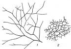
According to Krasilnikov’s classification, built taking into account the morphological structure and phylogenetic relationships of radiant fungi, the genus Actinomyces unites organisms with well-developed, nonseptate mycelium, which does not break up into rod-shaped and coccoid elements. Since there are no partitions in the mycelial threads, they represent one cell (Fig. 1). On agar media, different types of A. form different colonies: flat, wrinkled, smooth, tuberous, filmy, etc. Substrate mycelium extends from the lower surface of the colonies and grows into the medium; from the surface of the colonies among the aerial mycelium, spore carriers develop - straight or spirally twisted, with the number of curls up to 10 or more (Fig. 2). Spores are formed within the spore bearers by fragmentation or segmentation. In the first case, the separation of individual lumps of protoplasm inside the filament occurs, from which chains of spores are then formed. In the second, this process is preceded by segmentation of mycelial filaments. The spores are spherical, oval, rod-shaped, with a smooth or spiny surface (Fig. 3).

Different in chemistry. The composition of the pigments determines the different colors of the colonies (color table, article 184, Fig. 1-12). Different combinations of these pigments in the same colony create many different shades. Some pigments are soluble in water, others (for example, red-yellow lipoactinochromes) are soluble only in alcohol or other fat solvents. Some pigments remain in cells, others diffuse into the environment.
Most A. are aerobes; anaerobic or microaerophilic species are rare. A. growth occurs at a wide pH value from 5 to 9. The temperature optimum for most species is 25-30° (mesophiles), but growth is possible in the range from 3 to 40°. The thermophiles present among A. grow at t° 45-50°.
The presence of a large number of different enzymes in A. - proteases, keratinases, chitinases, lipases, amylase, invertase, etc. - increases A.'s ability to use plant and animal residues, as well as such substrates that other microorganisms do not use - paraffin, kerosene , wax, resin, etc. Some types of A. fix molecular nitrogen. Enzymatic activity is also manifested in the lytic processes characteristic of A., for example. autolysis, which can also be caused by a lytic effect on other microorganisms.
Many metabolites of A. belong to biologically active compounds - antibiotics, hormones, vitamins, enzymes, etc. Approximately. 1000 antibiotic substances active against bacteria, fungi, viruses, protozoa, and tumors. Many of them have received wide practical use - streptomycin, aureomycin, terramycin, etc. Certain A. toxins also have an antimicrobial effect, for example, gliotoxin, which is highly toxic to animals and plants.
Almost all A. are capable of synthesizing vitamin B 12 and its analogues. Some species synthesize vitamins Bx, B2, biotin, pantothenic and nicotinic acids, pyridoxine, riboflavin, etc. Some A. are producers of amino acids - glutamic, aspartic, valine, methionine, cysteine, cystine, etc. Some species produce aromatic substances with the odors of earth (the most characteristic feature of A.), fruit, camphor, iodoform, hydrogen sulfide, ammonia, etc. A. are widespread throughout the entire surface of the globe: there are many of them on plant and animal remains, in the water of natural reservoirs, and especially in the soil. From 1 G Soils are sown from several hundred to millions and even billions of actinomycetes, less from primary soils, more from cultivated soils.

A. widely participate in the cycle of substances in nature, breaking down many substrates inaccessible to other microorganisms, contributing to the formation of humus and weathering of rocks.
Waxman and Henrizi classify radiant fungi based on their pathogenicity, anaerobiosis, acid resistance and other characteristics. They retained the name of the genus Actinomyces as a historical name for pathogenic species. Vaksman and Henritsp included in the genus A. such radiant fungi, the body of which consists of a thin but septate mycelium, which disintegrates over time into rod-shaped and coccoid elements. These organisms are pathogenic, non-acid-fast, gram-positive, anaerobic. Colonies are leathery, dense or pasty, sometimes covered with sparse aerial mycelium. Representatives of this genus cause in humans and animals actinomycosis(cm.).
There are many types of A. that cause this disease, but the following types are more common. Actinomyces Israeli Lachner-Sandoval (1898); syn.: A. hominis Wolff-Israel (1891), Streptothrix israeli Kruss (1896), Proactinomyces israeli (Kruse) Krassilnikov (1941). Colonies are colorless, pasty, smooth, sometimes lumpy; aerial mycelium is represented by sparse branches and does not form pigments. It grows poorly on C synthetic media; it grows better on protein media with blood serum, in microaerophilic conditions at t° 37°. Microscopically it consists of thin filaments of mycelium (3-10×0.6 mk), disintegrating over time into polymorphic - rod-shaped, coccoid, flask-shaped, spherical, spindle-shaped - elements. Assimilates glucose, maltose, sucrose, galactose, fructose, lactose, mannose, raffinose and other sugars. It does not liquefy gelatins, does not peptonize or curdle milk, does not restore nitrates, and weakly hydrolyzes starch. Pathogenic for humans and certain animals.
Actinomyces bovis Harz (1877); syn.: Discomyces bovis Rivolta (1878), Cladothrix bovis Mace (1891), Nocardia actinomyces Trevisan (1889), Proactinomyces bovis (Wright) Krassilnikov (1941). Colonies are colorless, dough-like, sometimes leathery, covered with white aerial mycelium, which breaks down into rod-shaped and coccoid elements. The spore bearers on the aerial mycelium are slightly wavy, but not spiral. Anaerobe. Grows well on protein media with t° 37°. Does not liquefy gelatins, does not ferment milk, does not hydrolyze starch. Assimilates glucose, galactose, fructose, mannose, glycerol; weaker - maltose, sucrose, inulin, mannitol, dulcite, lactose. Under natural conditions it affects cattle, horses, pigs and other animals, and also occurs in humans.
Organisms of the same structure, but growing under aerobic conditions and partially acid-resistant, were identified by Waxman and Henrizi as a special genus Nocardia, whose representatives cause disease in humans and animals - nocardiosis(cm.).
Pathogenic species of A. live in the surrounding nature, but are found as saprophytes in the body of people and animals, Ch. arr. in the oral cavity (in tartar, dental plaque), so infection with actinomycosis can be both exogenous and endogenous.
The genus Proactinomyces, or Nocardia (according to Krasilnikov), includes two genera - Actinomyces and Nocardia Waksman a. Henrici, as well as numerous saprophytic species of the same structure.
Among the causative agents of actinomycosis there are representatives of the genus Micromonospora, which is also included in the extensive class of radiant fungi Actinomycetes, family. Micromonosporaceae (according to Krasilnikov) or Streptomycetaceae (according to Vaksman and Henrizi). They cause disease - micromonosporosis(cm.). The structure of micromonospores is similar to that of Actinomyces and Proactinomyces. The difference lies in the method of formation of spores (conidia), which are formed one at a time at the end of the spore carrier. In nature, they are less common than A. Of the pathogenic species, the most common is Micromonospora parva Jensen (1932). The colonies are flat, bare, with rare conidiophores, at the ends of which there is one oval spore on a stalk or directly on the mycelial thread; These spores in bulk have a grayish-greenish color. The orange pigment of the colonies themselves does not diffuse into the medium. Mesophiles. Aerobes. Chemically inactive: gelatin is slightly liquefied, milk is not changed, starch is hydrolyzed.
Micromonospora monospora (Lehmann, Schitze) Jensen (1932); synonym: Actinomyces monosporus Lehmann, Schutze (1908), Thermoactinomyces monosporus Waksman (1961). The colonies are covered with aerial mycelium, the conidiophores bear one oval spore. The color of the colonies is yellow or gray-green. Temperature optimum is 37°, can withstand short-term heating up to 55-75° and higher. The gelatin is liquefied, the milk is not changed. Aerophiles.
S. F. Dmitriev also described the phenomenon of spontaneous lysis, widespread among pathogenic A. This property of A. is used to obtain the drug actinolysate, which in the USSR is used for the treatment and diagnosis of actinomycosis.
Z. G. Stepanishcheva.
Sources:
- Big medical encyclopedia. Volume 1/Editor-in-Chief Academician B.V. Petrovsky; publishing house "Soviet Encyclopedia"; Moscow, 1974.- 576 p.
- 1.Medical microbiology. Subject, tasks, methods, connection with other sciences. The importance of medical microbiology in the practical activities of a doctor.
- 3. Microorganisms and their position in the system of the living world. Nomenclature of bacteria. Principles of classification.
- 6. Growth and reproduction of bacteria. Reproduction phases.
- 7. Nutrition of bacteria. Types and mechanisms of bacterial nutrition. Autotrophs and heterotrophs. Growth factors. Prototrophs and auxotrophs.
- 8. Nutrient media. Artificial nutrient media: simple, complex, general purpose, elective, differential diagnostic.
- 9. Bacteriological method of studying microorganisms. Principles and methods for isolating pure cultures of aerobic and anaerobic bacteria. The nature of the growth of microorganisms on liquid and solid nutrient media.
- 13. Spirochetes, their morphology and biological properties. Species pathogenic to humans.
- 14. Rickettsia, their morphology and biological properties. The role of rickettsia in infectious pathology.
- 15. Morphology and ultrastructure of mycoplasmas. Species pathogenic to humans.
- 16. Chlamydia, morphology and other biological properties. Role in pathology.
- 17. Fungi, their morphology and biological features. Principles of taxonomy. Diseases caused by fungi in humans.
- 20. Interaction of virus with cell. Life cycle phases. The concept of persistence of viruses and persistent infections.
- 21. Principles and methods of laboratory diagnosis of viral infections. Virus cultivation methods.
- 24. Structure of the bacterial genome. Mobile genetic elements, their role in the evolution of bacteria. The concept of genotype and phenotype. Types of variability: phenotypic and genotypic.
- 25. Bacterial plasmids, their functions and properties. Use of plasmids in genetic engineering.
- 26. Genetic recombinations: transformation, transduction, conjugation.
- 27. Genetic engineering. The use of genetic engineering methods to obtain diagnostic, preventive and therapeutic drugs.
- 28.Distribution of microbes in nature. Microflora of soil, water, air, methods of studying it. Characteristics of sanitary indicator microorganisms.
- 29. Normal microflora of the human body, its role in physiological processes and pathology. The concept of dysbacteriosis. Preparations for restoring normal microflora: eubiotics (probiotics).
- 31. Forms of manifestation of infection. Persistence of bacteria and viruses. The concept of relapse, reinfection, superinfection.
- 32. Dynamics of development of the infectious process, its periods.
- 33. The role of microorganisms in the infectious process. Pathogenicity and virulence. Units of measurement of virulence. The concept of pathogenicity factors.
- 34. Classification of pathogenicity factors according to o.V. Bukharin. Characteristics of pathogenicity factors.
- 35. The concept of immunity. Types of immunity.
- 36. Nonspecific protective factors of the body against infection. Role of I.I. Mechnikov in the formation of the cellular theory of immunity.
- 37. Antigens: definition, basic properties. Antigens of bacterial cells. Practical use of bacterial antigens.
- 38. Structure and functions of the immune system. Cooperation of immunocompetent cells. Forms of immune response.
- 39. Immunoglobulins, their molecular structure and properties. Immunoglobulin classes. Primary and secondary immune response. :
- 40. Classification of hypersensitivity according to Jail and Coombs. Stages of an allergic reaction.
- 41. Immediate hypersensitivity. Mechanisms of occurrence, clinical significance.
- 42. Anaphylactic shock and serum sickness. Causes of occurrence. Mechanism. Their warning.
- 43. Delayed hypersensitivity. Skin allergy tests and their use in the diagnosis of certain infectious diseases.
- 44. Features of antiviral, antifungal, antitumor, transplantation immunity.
- 45. Concept of clinical immunology. Human immune status and factors influencing it. Assessment of immune status: main indicators and methods for their determination.
- 46. Primary and secondary immunodeficiencies.
- 47. Interaction of antigen with antibody in vitro. Theory of network structures.
- 48. Agglutination reaction. Components, mechanism, installation methods. Application.
- 49. Coombs reaction. Mechanism. Components. Application.
- 50. Passive hemagglutination reaction. Mechanism. Components. Application.
- 51. Hemagglutination inhibition reaction. Mechanism. Components. Application.
- 53. Complement fixation reaction. Mechanism. Components. Application.
- 54. The reaction of neutralizing a toxin with an antitoxin, neutralizing viruses in cell culture and in the body of laboratory animals. Mechanism. Components. Staging methods. Application.
- 55. Immunofluorescence reaction. Mechanism. Components. Application.
- 56. Enzyme immunoassay. Immunoblotting. Mechanisms. Components. Application.
- 57. Vaccines. Definition. Modern classification of vaccines. Requirements for vaccine products.
- 59. Vaccine prevention. Vaccines made from killed bacteria and viruses. Cooking principles. Examples of killed vaccines. Associated vaccines. Advantages and disadvantages of killed vaccines.
- 60. Molecular vaccines: toxoids. Receipt. Use of toxoids for the prevention of infectious diseases. Examples of vaccines.
- 61. Genetically engineered vaccines. Receipt. Application. Advantages and disadvantages.
- 62. Vaccine therapy. The concept of therapeutic vaccines. Receipt. Application. Mechanism of action.
- 63. Diagnostic antigenic preparations: diagnosticums, allergens, toxins. Receipt. Application.
- 64. Serums. Definition. Modern classification of serums. Requirements for whey preparations.
- 65. Antibody preparations are serums used for the treatment and prevention of infectious diseases. Methods of obtaining. Complications during use and their prevention.
- 66. Antibody preparations are sera used to diagnose infectious diseases. Methods of obtaining. Application.
- 67. Concept of immunomodulators. Operating principle. Application.
- 68. Interferons. Nature, methods of production. Application. No. 99 Interferons. Nature, methods of production. Application.
- 69. Chemotherapy drugs. The concept of the chemotherapeutic index. The main groups of chemotherapeutic drugs, the mechanism of their antibacterial action.
- 71. Drug resistance of microorganisms and the mechanism of its occurrence. The concept of hospital strains of microorganisms. Ways to overcome drug resistance.
- 72. Methods for microbiological diagnosis of infectious diseases.
- 73. Causative agents of typhoid fever and paratyphoid fever. Taxonomy. Characteristic. Microbiological diagnostics. Specific prevention and treatment.
- 74. Pathogens of escherichiosis. Taxonomy. Characteristic. The role of Escherichia coli in normal and pathological conditions. Microbiological diagnosis of escherichiosis.
- 75. Pathogens of shigellosis. Taxonomy. Characteristic. Microbiological diagnostics. Specific prevention and treatment.
- 76. Pathogens of salmonellosis. Taxonomy. Characteristics. Microbiological diagnosis of salmonellosis. Treatment.
- 77. Pathogens of cholera. Taxonomy. Characteristic. Microbiological diagnostics. Specific prevention and treatment.
- 78. Staphylococci. Taxonomy. Characteristic. Microbiological diagnosis of diseases caused by staphylococci. Specific prevention and treatment.
- 79. Streptococci. Taxonomy. Characteristic. Microbiological diagnosis of streptococcal infections. Treatment.
- 80. Meningococci. Taxonomy. Characteristic. Microbiological diagnosis of streptococcal infections. Treatment.
- 81. Gonococci. Taxonomy. Characteristic. Microbiological diagnosis of gonorrhea. Treatment.
- 82. Causative agent of tularemia. Taxonomy. Characteristics. Microbiological diagnostics. Specific prevention and treatment.
- 83. The causative agent of anthrax. Taxonomy and characteristics. Microbiological diagnostics. Specific prevention and treatment.
- 84. Causative agent of brucellosis. Taxonomy and characteristics. Microbiological diagnostics. Specific prevention and treatment.
- 85. Causative agent of plague. Taxonomy and characteristics. Microbiological diagnostics. Specific prevention and treatment.
- 86. Pathogens of anaerobic gas infection. Taxonomy and characteristics. Microbiological diagnostics. Specific prevention and treatment.
- 87. Causative agents of botulism. Taxonomy and characteristics Microbiological diagnostics. Specific prevention and treatment.
- 88. The causative agent of tetanus. Taxonomy and characteristics. Microbiological diagnosis and treatment.
- 89. Non-spore-forming anaerobes. Taxonomy. Characteristics. Microbiological diagnosis and treatment.
- 90. The causative agent of diphtheria. Taxonomy and characteristics. Conditionally pathogenic corynebacteria. Microbiological diagnostics. Detection of anoxic immunity. Specific prevention and treatment.
- 91. Pathogens of whooping cough and parawhooping cough. Taxonomy and characteristics. Microbiological diagnostics. Specific prevention and treatment.
- 92. Pathogens of tuberculosis. Taxonomy and characteristics. Conditionally pathogenic mycobacteria. Microbiological diagnosis of tuberculosis.
- 93. Actinomycetes. Taxonomy. Characteristic. Microbiological diagnostics. Treatment.
- 95. The causative agent of chlamydia. Taxonomy. Characteristics. Microbiological diagnostics. Treatment.
- 96. Causative agent of syphilis. Taxonomy. Characteristic. Microbiological diagnostics. Treatment.
- 97. Causative agent of leptospirosis. Taxonomy. Characteristics. Microbiological diagnostics. Specific prevention. Treatment.
- 98. Causative agent of borreliosis. Taxonomy. Characteristics. Microbiological diagnostics.
- 99. Clinical microbiology, its tasks. Vbi, features of the cause of occurrence. The role of conditionally pathogenic microorganisms in the occurrence of nosocomial infections.
- 100. Classification of mushrooms. Characteristic. Role in pathology. Laboratory diagnostics. Treatment.
- 101. Classification of mycoses. Superficial and deep mycoses. Yeast-like fungi of the genus Candida. Role in human pathology.
- 102. The causative agent of influenza. Taxonomy. Characteristic. Laboratory diagnostics. Specific prevention and treatment.
- 103. The causative agent of polio. Taxonomy and characteristics. Laboratory diagnostics. Specific prevention.
- 104. Pathogens of hepatitis a and e. Taxonomy. Characteristics. Laboratory diagnostics. Specific prevention.
- 105. Causative agent of tick-borne encephalitis. Taxonomy. Characteristics. Laboratory diagnostics. Specific prevention.
- 106. Rabies agent. Taxonomy. Characteristics. Laboratory diagnostics. Specific prevention.
- 107. The causative agent of rubella. Taxonomy. Characteristic. Laboratory diagnostics. Specific prevention.
- 108. Measles virus. Taxonomy. Characteristic. Laboratory diagnostics. Specific prevention.
- 109. Causative agent of mumps. Taxonomy. Characteristic. Laboratory diagnostics. Specific prevention.
- V.Clinic
- I. Epidemiology
93. Actinomycetes. Taxonomy. Characteristic. Microbiological diagnostics. Treatment.
Morphology. Genus Actynomyces Branching bacteria. They do not contain chitin in the cell wall; the wall has the structure of gram-positive bacteria. The mycelium looks like thin straight rods, forming threads. A characteristic feature of actinomycetes is the ability to form well-developed mycelium. Rod-shaped forms, often with thickened ends, are arranged singly, in pairs, in V- and Y-shapes in the smear. All morphological forms are capable of true branching, especially in a thioglycollate semi-liquid medium. Gram staining is poor, often forming granular or clear-shaped forms; non-acid-resistant. Typical view - Actinomyces bovis.
Cultural properties. Obligate and facultative anaerobes. They grow slowly, crops should be cultivated for 7 days. Temperature optimum for growth is 37C. Some strains produce α-β-hemolysis on blood media. Some species form filamentous microcolonies resembling mycelium; on the 7th day they form S-shaped colonies, sometimes colored yellow/red. A. odontolyticus On blood agar it forms red colonies with a zone of β-hemolysis.
Biochemical activity. Chemoorganotrophs. Carbohydrates are fermented to form acid without gas; fermentation products are acetic, formic, lactic and succinic acids. The presence of catalase and the ability to reduce nitrates into nitrites do not form indole.
Antigenic structure. ELISA distinguishes 6 groups: A, B, C, D, E and F.
Antimicrobial sensitivity. Sensitive to penicillins, tetracycline, erythromycin, but resistant to antimycotics. Sensitive to the effects of commonly used antiseptics and disinfectants.
Epidemiology. The source of infection is soil. The transmission mechanism is contact, and the transmission route is wound. They colonize the oral mucosa of humans and mammals.
Pathogenesis. Causes opportunistic infection.
Clinic. Actinomycosis is a chronic opportunistic infection of humans and animals caused by anaerobic and facultative anaerobic actinomycetes, which is characterized by granulomatous inflammation.
: Material for research - sputum, cerebrospinal fluid, pus from fistulas, tissue biopsy.
Used for diagnosis bacterioscopic, bacteriological, serological And allergological methods.
Bacteroscopically: by detecting drusen of actinomycetes in the test material, which look like small yellowish or grayish-white grains with a greenish tint. According to Gram, spores are colored dark purple, mycelium is purple, and drusen are pink.
To suppress the growth of accompanying microflora, pus and sputum are centrifuged in a solution of penicillin and streptomycin before inoculation. Inoculated on nutrient media (sugar agar) and cultivated under aerobic and anaerobic conditions. The ability of isolated cultures to curdle and peptonize milk is determined - a characteristic characteristic of actinomycetes. Isolation of anaerobic species confirms the diagnosis of actinomycosis.
For serodiagnosis, RSC with actinolysate is performed. The reaction is not specific enough, since positive results can be observed with lung cancer and severe suppurative processes. The use of actinomycetes as an antigen instead of actinolysate increases the sensitivity of CSCs. The same AG can also be used to stage RIGA.
An allergy test is carried out with actinolysate. Positive tests have diagnostic value.
Treatment. The use of penicillin, tetracycline, erythromycin, clindamycin.
Prevention. Specific prevention - no. Nonspecific - increased immune status.
94. Pathogens of rickettsiosis. Taxonomy. Characteristic. Microbiological diagnostics. Specific prevention and treatment (typhus) Epidemic typhus - acute anthroponosis with a transmissible mechanism of spread by body lice. Clinically characterized by fever, severe course due to damage to the blood capillaries with impaired blood supply to vital organs (brain, heart, kidneys), and the appearance of a rash.
Epidemiology and mechanism of infection. Infection occurs either by rubbing feces of infected lice through scratching the skin, or by inhaling a dusty aerosol from dried feces infected with rickettsia.
Clinic, diagnosis, treatment. The incubation period is 10 days. The onset of the disease is acute, clinical manifestations are caused by generalized damage to the system of endothelial cells of blood vessels, which leads to disruption of the thrombo-antithrombotic cascade. The morphological basis of the disease is generalized vasculitis with the formation of a rash on the skin. The disease occurs with high fever, symptoms of damage to the cardiovascular and nervous systems. Immunity- short-lived, cellular-humoral.
Diagnostics: carried out according to clinical and epidemiological data, supported by laboratory testing for specific antibodies (RSK, RNGA, ELISA, etc.).
Treatment: Rapid etiotropic treatment with a single dose of doxycycline, or in its absence - with tetracycline drugs.
Prevention. Isolation of lice-infested patients, disinfection with preparations containing permethrin. For specific prevention, a live vaccine from the E strain has been developed, which is used in combination with the soluble Provacek rickettsia antigen (live combined typhus vaccine from the strain), as well as an inactivated vaccine from the soluble antigen.
Brill's disease – relapse after previously suffering epidemic typhus.
Pathogen- R. prowazekii.
Clinically proceeds as epidemic typhus of mild to moderate severity.
Pathomorphology infectious process is the same as in the epidemic form. The difference lies in epidemiology (no carrier, no seasonality of manifestation, source and implementation of the method of infection) and pathogenesis of the initial stage of the disease. It occurs due to the activation of latent “dormant” rickettsiae.
Microbiological diagnostics . It is complicated by the uncertainty of symptoms in the first week of the disease (before the appearance of the rash) and its similarity to the symptoms of infections, most often typhoid. The diagnosis is established on the basis of clinical and epidemiological data, taking into account the patient’s medical history and is supported by a serological test with a specific antigen. In the absence of a carrier in the outbreak, treatment can be carried out without isolating the patient, depending on his condition. The prognosis is favorable even in the absence of antibiotic treatment.
Prevention. Prevention measures are the same as for the epidemic form. Specific prevention is not possible.
Many researchers (Krasilnikov, 1950; Erikson, 1953; Prevot, 1953; etc.) noted their similarity to fungi and considered them as an intermediate form between fungi and bacteria.
The study of the ultrastructural organization of actinomycete cells, as well as the analysis of chemical components, made it possible to classify them as belonging to the kingdom of prokaryotes and consider them one of the groups of bacteria “in the broad sense” (Murray, 1960; Stanier, 1962). With this understanding, the group of bacteria includes almost all prokaryotes (rickettsia and mycoplasmas, blue-green algae, cyanobacteria and mycobacteria).
Actinomycetes- a peculiar prokaryotic group of microorganisms, their cells form branching outgrowths, and some have mycelium 0.5-2 microns thick, usually appearing in extreme conditions. The cell wall is similar to that of gram-positive bacteria. The cell contains a nucleoid, vacuoles, volutin, and mesosomes in the cytoplasmic membrane. Developing in surface cultures, they form two-phase mycelium - in the substrate and on the surface (the latter is differentiated). Actinomycetes reproduce by fragments of mycelium or spores. Spores are formed asexually on aerial or substrate mycelium and are located singly, in pairs, in chains, or concentrated in sporangia. Some of them are movable. Based on the type of sporogenesis and morphological characteristics, the class Actinomycetes is divided into orders, families and genera. All actinomycetes are heterotrophs and are usually not demanding on nutrition. In relation to oxygen, they are mainly aerobes, although anaerobes are also found among pathogenic ones. They usually develop in a neutral reaction environment, but there are also acidophilic forms (acid-resistant) and alkali-resistant, many of them can withstand drying and salinity.
Actinomycetes are widely distributed in the air, water bodies, plant and animal remains, soils, especially in low-fertility soils - dry, desert, saline, rocky, which are in the first stages of the soil-forming process. They participate in the transformation of organic and mineral substances and are producers of many physiologically active substances, enzymes (chitinases, etc.) - amino acids, vitamins, antibiotics, and enrich the soil with nitrogen.
Coryne-like bacteria are related to actinomycetes. This group includes Corynebacterium, Arthrobacter and other genera found in various types of soils and rocks. The genus Corynebacterium is represented by bacterial forms that are prone to variability (in the form of rods, cocci, branching forms); daughter cells are often located at an angle or adjacent to each other. All of them are chemoorganotrophs, obligate aerobes, but there are also facultative aerobes. Bacteria of the genus Arthrobacter are the main representatives of soil microflora, actively participating in the decomposition of organic matter, characterized by rod-shaped or coccoid-shaped cells and tend to branch; are obligate aerobic chemoorganoheterotrophs.
According to Bergi's determinant, the department Bacteria also includes mycoplasmas, which play an important role in the cycle of Fe and Mn. These microorganisms do not have a cell wall, they are characterized by polymorphism, they reproduce by division and fragmentation of large bodies. The sizes of the largest mycoplasma cells reach 10 microns, the majority - 0.1-0.2 microns. A nucleotide with DNA was found inside the cell, and ribosomes were found in the cytoplasm; there are no mesosomes. Among them there are aerobes and anaerobes, acidophiles and thermophiles, but all of them are insensitive to antibiotics due to the lack of a cell membrane. Mycoplasmas are found in soils, reservoirs, hot springs and on plants. Many of them are pathogens of diseases in humans, animals and plants.
Actinomycetes (aeum - ray, tybm - fungus) - radiant fungi, represent a large group of microorganisms included in the order Acidomycetes,
Most actinomycetes are free-living microorganisms that live in soil and other environmental objects. Many of them are producers of antibiotics. A few pathogenic representatives of actinomycetes cause actinomycosis and nocardiosis in humans.
Actinomycete cells have the same structural elements as bacteria: cell wall, CPM; the cytoplasm contains a nucleoid, ribosomes, mesosomes, and intracellular inclusions (Fig. 13). Some actinomycetes form a microcapsule.
The main morphological feature of actinomycetes is the branching form of cells that look like short rods or long filamentous formations, reminiscent of fungal mycelium and therefore called hyphae. Mycelium can be substrate (growing into a dense medium) or aerial.
The width of actinomycete cells is 0.2-0.5 microns, the length can vary widely. In the body of people and animals with actinomycosis, pathogenic actinomycetes form peculiar accumulations of altered mycelium - drusen.
Actinomycetes differ from each other in the structure of the peptidoglycan layer of the cell wall. Peptidoglycan peptides in most species contain the same four amino acids found in bacteria. Unlike bacteria, the peptidoglycan of some actinomycetes contains sugars such as arabinose, galactose, xylose, and madurose. The morphology of actinomycetes is studied in stained smears and using phase-contrast microscopy, as well as electron microscopy.
Actinomycosis is an opportunistic infection caused by actinomycetes and characterized by granulomatous inflammation with polymorphic clinical manifestations.
The formation of a granuloma undergoing necrotic decay with the formation of pus emerging through fistulas onto the surface of the skin and mucous membranes. Pus of varying consistency, sometimes mixed with blood, often contains drusen. At the same time, granuloma fibrosis is noted. Depending on the location: cervicofacial, thoracic, abdominal, genitourinary, osteoarticular, musculocutaneous, septic and other forms.
For example, actinomycetes belonging to the species Actinomyces streptomycin! inhibit the growth of gram-positive and gram-negative bacteria, mycobacteria, and some types of yeast and fungi. Actinomyces levoris does not inhibit the growth of bacteria, but inhibits the development of yeast, some yeast-like organisms, filamentous fungi, etc. The antimicrobial spectrum of action is one of the taxonomic characteristics in the taxonomy of actinomycetes that serve to distinguish species. Antibiotics produced by actinomycetes do not inhibit the development of their own culture, even in concentrations that are many times higher than the minimum concentration that inhibits the growth of other microorganisms.
From a culture of the purple actinomycete Actinomyces violaceus, which they isolated from the soil, they obtained the first antibiotic of actinomycete origin - mycetin - and studied the conditions for the biosynthesis and use of mycetin in the clinic.
The antibiotic actinomycin was isolated from the culture of Actinomyces antibioticus, which subsequently began to be used as an anticancer agent. The first antibiotic of actinomycete origin, which found widespread use especially in the treatment of tuberculosis, was streptomycin, discovered in 1944 by Waksman and co-workers. Anti-tuberculosis antibiotics also include the later discovered viomycin (florimycin), cycloserine, kanamycin, and rifamycin.
18. Spirochetes, their morphology and biological properties
Spirochetes (bacteria in the form of a curved long screw) differ from bacteria in structure. They have a corkscrew, twisted shape. Their sizes vary widely (width 0.3-1.5 microns and length 7-500 microns). The body of the spirochete consists of an axial filament and cytoplasm, spirally curled around the filament. Spirochetes have a three-layer outer membrane. Electron microscopy revealed a delicate cytoplasmic membrane containing the cytoplasm. They do not form spores, capsules or flagella. In some species, an electron microscope reveals very thin thread-like fibril formations at the ends.
Spirochetes have active motility due to the pronounced flexibility of their body. In spirochetes, rotational, translational, wave-like, and bending movements are distinguished.
According to Romanovsky-Giemsa, some species are colored blue, others blue-violet, and others pink. A good method for processing spirochetes is silvering. Tinctorial properties are used to differentiate saprophytes and pathogenic spirochetes.
Three genera are pathogenic: Treponema, Borrelia, Leptospira.
Morphology. Genus Actynomyces Branching bacteria. They do not contain chitin in the cell wall; the wall has the structure of gram-positive bacteria. The mycelium looks like thin straight rods, forming threads. A characteristic feature of actinomycetes is the ability to form well-developed mycelium. Rod-shaped forms, often with thickened ends, are arranged singly, in pairs, in V- and Y-shapes in the smear. All morphological forms are capable of true branching, especially in a thioglycollate semi-liquid medium. Gram staining is poor, often forming granular or clear-shaped forms; non-acid-resistant. Typical view - Actinomyces bovis.
Cultural properties. Obligate and facultative anaerobes. They grow slowly, crops should be cultivated for 7 days. Temperature optimum for growth is 37C. Some strains produce α-β-hemolysis on blood media. Some species form filamentous microcolonies resembling mycelium; on the 7th day they form S-shaped colonies, sometimes colored yellow/red. A. odontolyticus On blood agar it forms red colonies with a zone of β-hemolysis.
Biochemical activity. Chemoorganotrophs. Carbohydrates are fermented to form acid without gas; fermentation products are acetic, formic, lactic and succinic acids. The presence of catalase and the ability to reduce nitrates into nitrites do not form indole.
Antigenic structure. ELISA distinguishes 6 groups: A, B, C, D, E and F.
Antimicrobial sensitivity. Sensitive to penicillins, tetracycline, erythromycin, but resistant to antimycotics. Sensitive to the effects of commonly used antiseptics and disinfectants.
Epidemiology. The source of infection is soil. The transmission mechanism is contact, and the transmission route is wound. They colonize the oral mucosa of humans and mammals.
Pathogenesis. Causes opportunistic infection.
Clinic. Actinomycosis is a chronic opportunistic infection of humans and animals caused by anaerobic and facultative anaerobic actinomycetes, which is characterized by granulomatous inflammation.
Microbiological diagnostics : Material for research - sputum, cerebrospinal fluid, pus from fistulas, tissue biopsy.
Used for diagnosis bacterioscopic, bacteriological, serological And allergological methods.
Bacteroscopically: by detecting drusen of actinomycetes in the test material, which look like small yellowish or grayish-white grains with a greenish tint. According to Gram, spores are colored dark purple, mycelium is purple, and drusen are pink.
To suppress the growth of accompanying microflora, pus and sputum are centrifuged in a solution of penicillin and streptomycin before inoculation. Inoculated on nutrient media (sugar agar) and cultivated under aerobic and anaerobic conditions. The ability of isolated cultures to curdle and peptonize milk is determined - a characteristic characteristic of actinomycetes. Isolation of anaerobic species confirms the diagnosis of actinomycosis.
For serodiagnosis, RSC with actinolysate is performed. The reaction is not specific enough, since positive results can be observed with lung cancer and severe suppurative processes. The use of actinomycetes as an antigen instead of actinolysate increases the sensitivity of CSCs. The same AG can also be used to stage RIGA.
An allergy test is carried out with actinolysate. Positive tests have diagnostic value.
Treatment. The use of penicillin, tetracycline, erythromycin, clindamycin.
Prevention. Specific prevention - no. Nonspecific - increased immune status.