Physicochemical methods of analysis (PCMA) are based on the use of the relationship between the measured physical properties of substances and their qualitative and quantitative composition. Since the physical properties of substances are measured using various devices - “instruments”, these methods of analysis are also called instrumental methods.
The greatest practical applications among FHMAs are:
- electrochemical methods– based on measuring potential, current, amount of electricity and other electrical parameters;
- spectral and other optical methods– based on the phenomena of absorption or emission of electromagnetic radiation (EMR) by atoms or molecules of a substance;
- chromatographic methods– are based on sorption processes occurring under dynamic conditions with directed movement of the mobile phase relative to the stationary one.
The advantages of PCMA include high sensitivity and low detection limit - mass up to 10-9 μg and concentration up to 10-12 g/ml, high selectivity (selectivity), which allows determining the components of mixtures without their preliminary separation, as well as rapid analysis, the possibility their automation and computerization.
Electrochemical methods are widely used in analytical chemistry. The choice of analysis method for a specific object of analysis is determined by many factors, including, first of all, the lower limit of the definition of the element.
Data on the lower limit of detection of various elements by some methods are presented in the table.
Limits of determination (μg/ml) of elements by various methods
| Element | MAS | AAS | PTP | WILLOW | Ionometry | Ampere.titles. |
| Ag | 0.1– dithizone | 0,07 | 0,2 | 0.00001 | 0.02 | 0.05 |
| As | 0.05 - molybd.blue | 0,2 | 0,04 | 0,02 | - | 0,05 |
| Au | 0.04-methyl.phiol. | 0,3 | 0,005 | 0,001 | - | 0,05 |
| Bi | 0.07-dithizone | 0,005 | 0,00001 | - | 0,5 | |
| Cd | 0.04-dithizone | 0,05 | 0,002 | 0,00001 | 0,03 | 0,5 |
| Cr | 0.04-diphenylcarbazide | 0,2 | 0,02 | - | - | |
| Cu | 0.03-dithizone | 0,2 | 0,002 | 0,00002 | 0,01 | 0,05 |
| Hg | 0.08-dithizone | - | 0,00005 | |||
| Pb | 0.08-dithizone | 0,6 | 0,003 | 0,00002 | 0,03 | |
| Sb | 0.08-rhodamine | 0,004 | 0,00004 | - | 0,5 | |
| Fe | 0,1-rhodanide | 0,2 | 0,003 | 0,0002 | 0,3 | 0,5 |
| Se | 0.08-diami-nophthalene | 0,3 | 0,2 | 0,00002 | - | 0,5 |
| Sn | 0.07-phenyl-fluriome | 0,4 | 0,003 | 0,00004 | - | 0,5 |
| Te | 0.1-bismuthol | 0,7 | 0,02 | - | - | |
| Tl | 0.06-rhodamine | 0,6 | 0,01 | 0,00002 | - | 0,5 |
| Zn | 0.02-dithizone | 0,02 | 0,003 | 0,0003 | - | 0,5 |
| F- | - | - | - | - | 0,02 | 5-10 |
| NH 4 + ,NO 3 - | - | - | - | - | 0,1 | 1-5 |
MAS - molecular absorption spectrometry (photometry);
AAS - atomic absorption spectrometry (flame photometry);
PTP - alternating current polarography;
IVA - stripping voltammetry.
Determination errors in FHMA are about 2-5%; carrying out analyzes requires the use of complex and expensive equipment.
Distinguish direct and indirect methods of physical and chemical analysis. Direct methods use the dependence of the magnitude of the measured analytical signal on the concentration of the component being determined. In indirect methods, the analytical signal is measured in order to find the end point of titration of the component being determined with a suitable titrant, that is, the dependence of the measured parameter on the volume of titrant is used.
Electrochemical methods of analysis are based on the study and use of processes occurring on the surface of the electrode or in the near-electrode space. Any electrical parameter (potential, electric current, amount of electricity, etc.), functionally related to the concentration of the component being determined and amenable to correct measurement, can serve as an analytical signal.
According to the nature of the measured analytical signal, electrochemical methods of analysis are divided into potentiometry, voltammetry, coulometry and a number of other methods:
Characteristic dependence of the electrochemical signal on the independent variable
| Method | Measured signal | Dependence of the signal on the independent variable |
| Potentiometry, ionometry | potential E = f(C) C-concentration of the analyte | |
| Potentiometric titration | potential E = f(V), V is the volume of the titrant reagent |
 |
| polarography, voltammetry | current I = f(E), E – electrode polarization potential |  |
| stripping voltammetry | current I n = f(E) |  |
| chronopotentiometry | potential E =f(t), t – electrode polarization time at I=const. |
 |
| amperometric titration with one indicator electrode | current I = f(V), V – volume of titrant reagent |  |
| amperometric titration with two indicator electrodes | current I = f(V) V – volume of titrant reagent | |
| coulometry | Q = f(C), C – amount of substance |  |
| conductometry | G = f(C), C – concentration of ions in solution |  |
| conductometric titration | electrical conductivity G = f(V), V – volume of titrant reagent |  |
Potentiometry
Potentiometric measurements are based on the dependence of the equilibrium potential of the electrode on the activity (concentration) of the ion being determined. For measurements, it is necessary to construct a galvanic cell from a suitable indicator electrode and reference electrode, and also have a device for measuring the potential of the indicator electrode (EMF of the galvanic cell), under conditions close to thermodynamic, when the indicator electrode has an equilibrium (or close to it) potential, that is, without removing noticeable current from the galvanic cell when the circuit is closed. In this case, you cannot use a regular voltmeter, but should use potentiometer- an electronic device with a high input resistance (1011 - 1012 Ohms), which eliminates the occurrence of electrode electrochemical reactions and the occurrence of current in the circuit.
An indicator electrode is an electrode whose potential depends on the activity (concentration) of the ion being determined in the analyzed solution.
A reference electrode is an electrode whose potential remains constant under analysis conditions. The potential of the indicator electrode is measured relative to the reference electrode. E(EMF of a galvanic cell).
In potentiometry, two main classes of indicator electrodes are used - electron exchange and ion exchange.
Electron exchange electrodes– these are electrodes on the surface of which electrode reactions involving electrons occur. Such electrodes include electrodes of the first and second kind, redox electrodes.
Electrodes of the first kind- these are electrodes that are reversible by a cation common to the electrode material, for example, metal M, immersed in a solution of a salt of the same metal. A reversible reaction M occurs on the surface of such an electrode n+ + ne↔ M and its real potential depends on the activity (concentration) of metal cations in solution in accordance with the Nernst equation:

For a temperature of 250C (298 K) and for conditions where the ion activity is approximately equal to the concentration (γ → 1):

Electrodes of the first kind can be made of various metals, for example, Ag (silver), Cu (copper), Zn (zinc), Pb (lead), etc.
Schematically, electrodes of the first kind are written as M | M n+ , where the vertical line shows the boundary of the solid (electrode) and liquid (solution) phases. For example, a silver electrode immersed in a solution of silver nitrate is depicted as follows - Ag | Ag+; if necessary, indicate the concentration of the electrolyte – Ag | AgNO 3 (0.1 M).
Electrodes of the first kind include gas hydrogen electrode Pt(H2) | H+ (2H + + 2e↔ H 2, E 0 = 0):

Electrodes of the second kind- these are anion-reversible electrodes, for example, a metal coated with a sparingly soluble salt of this metal, immersed in a solution containing the anion of this sparingly soluble salt M, MA | A n-. A reversible reaction MA + occurs on the surface of such an electrode ne↔ M + A n- and its real potential depends on the activity (concentration) of the anion in solution in accordance with the Nernst equation (at T= 298 K and γ → 1):

Examples of electrodes of the second type are silver chloride (AgCl + e↔ Ag + Cl -) and calomel (Hg 2 Cl 2 + 2e↔ 2Hg + 2Cl -) electrodes:

Redox electrodes– these are electrodes that consist of an inert material (platinum, gold, graphite, glassy carbon, etc.) immersed in a solution containing oxidized (Ok) and reduced (Boc) forms of the substance being determined. A reversible reaction Oc + occurs on the surface of such an electrode ne↔ Boc and its real potential depends on the activity (concentration) of the oxidized and reduced forms of the substance in solution in accordance with the Nernst equation (at T= 298 K and γ → 1):

If hydrogen ions participate in the electrode reaction, then their activity (concentration) is taken into account in the corresponding Nernst equations for each specific case.
Ion exchange electrodes- These are electrodes on the surface of which ion exchange reactions occur. Such electrodes are also called ion-selective or membrane. The most important component of such electrodes is semi-permeable membrane– a thin solid or liquid film that separates the inner part of the electrode (internal solution) from the analyzed solution and has the ability to transmit only ions of one type X (cations or anions). Structurally, the membrane electrode consists of an internal reference electrode (usually silver chloride) and an internal electrolyte solution with a constant concentration of the potential-determining ion, separated from the external (tested) solution by a sensitive membrane.
The real potential of ion-selective electrodes, measured relative to any reference electrode, depends on the activity of those ions in the solution that are sorbed by the membrane:

Where const – constant depending on the nature of the membrane ( asymmetry potential) and the potential difference between the external and internal reference electrodes, n And A(X n±) – charge and activity of the potential-determining ion. If the ion selective electrode potential is measured relative to a standard hydrogen electrode, then the constant is the standard electrode potential E 0.
For membrane electrodes the value slope of the electrode function may differ from theoretical Nernstovskaya magnitude (0.059 V); in this case, the real value of the electrode function is θ is determined as the tangent of the slope of the calibration graph. Then:

The potential of a membrane electrode in a solution containing, in addition to the detectable ion X, an extraneous ion B, which affects the potential of the electrode, is described Nikolsky equation(modified Nernst equation):

Where z– charge of foreign (interfering) ion, KХ/В – selectivity coefficient of the membrane electrode.
Selectivity coefficient K X/B characterizes the sensitivity of the electrode membrane to detectable X ions in the presence of interfering ions B. If K H/V<1, то электрод селективен относительно ионов Х и, чем меньше числовое значение коэффициента селективности, тем выше селективность электрода по отношению к определяемым ионам и меньше мешающее действие посторонних ионов. Если коэффициент селективности равен 0,01, то это означает, что мешающий ион В оказывает на величину электродного потенциала в 100 раз меньшее влияние, чем определяемый ион той же молярной концентрации.
The selectivity coefficient is calculated as the ratio of the activities (concentrations) of the detected and interfering ions, at which the electrode acquires the same potential in solutions of these substances, taking into account their charges:

Knowing the value of the selectivity coefficient, you can calculate the concentration of the interfering ion, which affects the potential of the ion-selective electrode (example).
Example. What concentration of nitrate ions must be created in a 1∙10-3 M sodium fluoride solution so that the ion-selective fluoride electrode is equally sensitive to both ions, if its electrode selectivity coefficient?

Solution.
Since then
This means that the concentration of nitrate ions in the analyzed solution above 0.5 mol/l has a significant effect on the determination of fluoride ion in its millimolar solutions.
A classic example of an ion-selective solid membrane electrode is a glass electrode with a hydrogen function, which is used to measure the concentration of hydrogen ions in a solution (glass pH electrode). For such electrodes, the membrane is a special glass of a certain composition, and the internal electrolyte is a 0.1 M solution of hydrochloric acid:
Ag, AgCl | 0.1 M HCl | glass membrane | test solution
An ion exchange process occurs on the surface of the glass membrane:
SiO-Na+ (glass) + H+ (solution) → -SiO-H+ (glass) + Na+ (solution)
as a result, a dynamic equilibrium is established between hydrogen ions in the glass and the solution H+ (glass) ↔ H+ (solution), which leads to the emergence of potential:
E = const + θ lg a(H+) = const – θ pH
A glass electrode with a high content of Al2O3 in the membrane measures the activity of sodium ions in solution (glass Na-electrode, sodium-selective electrode). In this case, the internal solution is 0.1 M sodium chloride solution:
Ag, AgCl | 0.1 M NaCl | glass membrane | test solution
On the surface of the glass membrane of the sodium-selective electrode, an equilibrium is established between sodium ions in the glass and the solution Na+ (glass) ↔ Na+ (solution), which leads to the emergence of potential:
E = const + θ lg a(Na+) = const – θ pNa
The most advanced electrode with a crystalline membrane is a fluoride-selective electrode, the membrane of which is made of a plate of lanthanum fluoride (LaF3) single crystal, activated to increase conductivity with europium fluoride (EuF 2):
Ag, AgCl | 0.1 M NaCl, 0.1 M NaF | LaF 3 (EuF 2) | test solution
The potential of the fluoride electrode is determined by the ion exchange process on its surface F- (membrane) ↔ F- (solution):
E = const – θ lg a(F-) = const + θ pF
Values of the constant and slope of the electrode function θ for ion-selective electrodes determined from the calibration graph E ÷рХ as a segment on the ordinate axis and the tangent of the angle of inclination of the straight line, respectively. For a glass pH electrode, this operation is replaced by setting up instruments (pH meters) using standard buffer solutions with precisely known pH values.
A schematic view of glass and fluoride-selective electrodes is shown in the figures:
Paired with an indicator electrode to measure its potential (EMF of a galvanic cell), a reference electrode with a known and stable potential, independent of the composition of the solution under study, is used. Silver chloride and calomel electrodes are most often used as reference electrodes. Both electrodes belong to the second type electrodes and are characterized by high stability in operation.
The potentials of silver chloride and calomel electrodes depend on the activity (concentration) of chloride ions (at T= 298 K and γ → 1):

Electrodes with a saturated solution of potassium chloride are most often used as reference electrodes - at 250C, the potential of a saturated silver chloride reference electrode is +0.201 V, and a saturated calomel electrode is +0.247 V (relative to the standard hydrogen electrode). Potentials for silver chloride and calomel reference electrodes containing 1 M and 0.1 M potassium chloride solutions can be found in the reference tables.
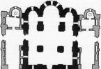
A schematic view of saturated silver chloride and calomel reference electrodes is shown in the figure:

Silver chloride reference electrodes (A) and calomel (b)
1 - asbestos fiber providing contact with the analyzed solution
2 - KCl solution (saturated)
3 - hole for contact
4 - solution of KCl (saturated), AgCl (solid)
5 - hole for introducing KCl solution
6 - paste from a mixture of Hg2Cl2, Hg and KS1 (saturated)
Potentiometric analysis is widely used to directly determine the activity (concentration) of ions in a solution by measuring the equilibrium potential of the indicator electrode (EMF of the galvanic cell) - direct potentiometry (ionometry), as well as to indicate the titration end point ( ktt) by changing the potential of the indicator electrode during the titration process ( potentiometric titration).
In all techniques direct potentiometry The dependence of the indicator electrode on the activity (concentration) of the ion being determined is used, which is described by the Nernst equation. The results of the analysis imply the determination of the concentration of a substance, and not its activity, which is possible when the ion activity coefficients are equal to unity (γ → 1) or their constant value (constant ionic strength of the solution), therefore, in further discussions only concentration dependences are used.
The concentration of the ion being determined can be calculated from the experimentally found potential of the indicator electrode if the constant component for the electrode is known (standard potential E 0) and the slope of the electrode function θ . In this case, a galvanic cell is composed, consisting of an indicator electrode and a reference electrode, its EMF is measured, the potential of the indicator electrode (relative to the SHE) and the concentration of the ion being determined are calculated.
IN method calibration chart prepare a series of standard solutions with a known concentration of the ion being determined and a constant ionic strength, measure the potential of the indicator electrode relative to the reference electrode (EMF of the galvanic cell) in these solutions and build a dependence based on the data obtained E÷ r WITH(A) (calibration graph). Then measure the potential of the indicator electrode in the analyzed solution E x (under the same conditions) and p is determined from the graph WITH x(A) and calculate the concentration of the analyte in the analyzed solution.
IN standard (comparison) method measure the potential of the indicator electrode in the analyzed solution ( E x) and in a standard solution of the analyte ( E st). The concentration of the ion being determined is calculated based on the Nernst equations for the analyzed sample and the standard sample. Slope of the electrode function for the indicator electrode θ
Using additive method first measure the potential of the indicator electrode in the analyzed solution ( E x), then add a certain volume of a standard solution of the analyte to it and measure the potential of the electrode in the resulting solution with the additive ( E x+d). The concentration of the ion being determined is calculated based on the Nernst equations for the analyzed sample and the sample with the additive. Slope of the electrode function for the indicator electrode θ must be known or determined in advance from the calibration schedule.
At potentiometric titration measure and record the EMF of the electrochemical cell (potential of the indicator electrode) after adding each portion of the titrant. Then, based on the results obtained, titration curves are constructed - integral in coordinates E ÷ V(a) And differential in coordinates ∆ E/∆V ÷ V (b), and determine the titration end point ( ktt) graphically:

Potentiometric titration uses all the main types of chemical reactions - acid-base, redox, precipitation and complexation. They are subject to the same requirements as in visual titrimetry, supplemented by the presence of a suitable indicator electrode to record changes in the concentration of potential-determining ions during titration.
The determination error during potentiometric titration is 0.5-1%, which is significantly lower than with direct potentiometric measurements (2-10%), however, higher detection limits are observed - more than 10 -4 mol/l.
Coulometry
Coulometry combines analysis methods based on measuring the amount of electricity spent on an electrochemical reaction. An electrochemical reaction results in quantitative electroconversion (oxidation or reduction) of the analyte at the working electrode (direct coulometry) or the production of an intermediate reagent (titrant) that reacts stoichiometrically with the analyte (indirect coulometry, coulometric titration).
The basis of coulometric methods is Faraday's law, which establishes a connection between the amount of electroconverted (oxidized or reduced) substance and the amount of electricity consumed:

Where m– mass of electroconverted substance, g; Q– the amount of electricity spent on the electrical transformation of a substance, C; F– Faraday number, equal to the amount of electricity required for the electrical transformation of one mole equivalent of a substance, 96500 C/mol; M– molar mass of the substance, g/mol; n– the number of electrons participating in the electrochemical reaction.
A necessary condition for carrying out coulometric analysis is the almost complete consumption of electricity for the transformation of the substance being determined, that is, the electrochemical reaction must proceed without side processes with 100% current efficiency.
In practice, coulometric analysis is implemented in two versions - at a constant potential ( potentiostatic coulometry) and at constant current ( amperostatic coulometry).
Potentiostatic coulometry used for direct coulometric measurements, when the substance being determined is directly subjected to electrolysis. In this case, the potential of the working electrode using potentiostats is maintained constant and its value is selected on the basis of polarization curves in the region of the limiting current of the substance being determined. During the electrolysis process at a constant potential, the current decreases in accordance with the decrease in the concentration of the electroactive substance according to the exponential law:

Where Ι – current strength at the moment of time t, A; Ι 0 – current strength at the initial moment of electrolysis, A; k– constant depending on electrolysis conditions.
Electrolysis is carried out until a residual current is reached Ι , the value of which is determined by the required accuracy - for a permissible error of 0.1%, electrolysis can be considered completed at Ι = 0,001Ι 0 . To reduce electrolysis time, a large surface working electrode should be used with intensive stirring of the analyzed solution.
Total amount of electricity Q, necessary for the electrical transformation of the analyte, is determined by the equation:

The amount of electricity can be determined by measuring the area under the current-time curve using mechanical or electronic integrators, or using chemical coulometers. Coulometer is an electrolytic cell in which an electrochemical reaction of known stoichiometry occurs with 100% current efficiency. The coulometer is connected in series with the coulometric cell under study, so during electrolysis the same amount of electricity flows through both cells. If, at the end of electrolysis, the amount (mass) of the substance formed in the coulometer is measured, then according to Faraday’s law, the amount of electricity can be calculated. The most commonly used are silver, copper and gas coulometers.
Amperostatic coulometry used for coulometric titration at a constant current, during which the analyte reacts with the titrant formed as a result of an electrochemical reaction on the working electrode, and therefore called electrogenerated titrant.
To ensure 100% current efficiency, a significant excess of the auxiliary substance from which the titrant is generated is required, which eliminates the occurrence of competing reactions at the working electrode. In this case, the titrant is generated in an amount equivalent to the analyte, and the amount of electricity spent on the generation of the titrant can be used to calculate the content of the analyte.
Amount of electricity Q in coulometry at constant current Ι calculated by the formula:

Where t– electrolysis time, for determining which almost all methods of establishing the end point in titrimetry are suitable (visual - indicators, instrumental - potentiometry, amperometry, photometry). With current in amperes and electrolysis time in seconds, we obtain the amount of electricity in coulombs (example).
Example. The coulometric titration of a solution of ascorbic acid with iodine generated from potassium iodide by a current of 5.00 mA took 8 min 40 s. Calculate the mass of ascorbic acid in the analyzed solution. Suggest a method for fixing the titration end point.
Solution. The amount of electricity spent on the oxidation of iodide and, accordingly, ascorbic acid is equal to:
Q = Ι·t= 5.00∙10 -3 ∙520 = 2.60 Cl.
Ascorbic acid is oxidized by iodine to dehydroascorbic acid with the release of two electrons (C 6 H 8 O 6 - 2 e→ C 6 H 6 O 6 + 2H +), then according to Faraday’s law:

The end point of the titration is determined by the appearance of excess iodine in the solution. Consequently, it can be detected visually using starch added to the analyzed solution (the appearance of a blue color), amperometrically with a dripping mercury or platinum microelectrode by the appearance of the limiting iodine current, or potentiometrically by a sharp increase in the potential of the platinum electrode.
Voltammetry
Voltammetric method of analysis is based on the use of the microelectrode polarization phenomenon, obtaining and interpreting current-voltage (polarization) curves reflecting the dependence of the current on the applied voltage. The current-voltage curve (voltammogram) allows you to simultaneously obtain qualitative and quantitative information about the substances that are reduced or oxidized on the microelectrode (depolarizers), as well as about the nature of the electrode process. Modern voltammetry is a highly sensitive and rapid method for determining substances, suitable for the analysis of various objects of inorganic and organic nature, including pharmaceuticals. The minimum detectable concentration in voltammetry reaches values of 10 -8 mol/l with a method error of less than 5%. Voltammetry under optimal experimental conditions makes it possible to determine several components simultaneously in the analyzed solution.
In voltammetry, two electrodes are used - worker a polarizable electrode with a small surface area (indicator microelectrode) and auxiliary non-polarizable electrode with a large surface area (reference electrode). The working electrodes are microelectrodes made of mercury (mercury dropping electrode, RCE), platinum (PE) and conductive carbon materials (graphite, glassy carbon).
When direct current passes through an electrolytic cell, the process is characterized by the relation (Ohm’s law for an electrolyte solution):
E = Ea – Ek + IR
Where E– applied external voltage; Ea– anode potential; Ek– cathode potential; I– current in the circuit; R– internal resistance of the electrolytic cell.
In voltammetric measurements, the analyzed solution contains an indifferent (background) electrolyte of high concentration (100 times or more higher than the concentration of the substance being analyzed - the solution resistance is low), and the current in voltammetry does not exceed 10 -5 A, therefore the voltage drop in the cell IR can be neglected.
Since in voltammetry one of the electrodes (auxiliary) is not polarized and the potential for it remains constant (it can be taken equal to zero), the voltage applied to the cell manifests itself in a change in the potential of only the working electrode and then E = Ea for the working microanode ( anodic polarization) And E =-Ek for the working microcathode ( cathodic polarization). Thus, the recorded current-voltage curve reflects the electrochemical process occurring only at the working electrode. If the solution contains substances that can be electrochemically reduced or oxidized, then when a linearly varying voltage is applied to the cell, the voltammogram has waveform 1 (in the absence of an electrochemical reaction, the dependence of the current on voltage is linear 2 in accordance with Ohm’s law):

The section of voltammetry in which the working microelectrode is an RCE is called polarography, in honor of the Czech electrochemist J. Heyrovsky, who proposed this method in 1922. Voltammograms obtained in a cell with a dropping mercury electrode are called polarograms.
To record classical polarograms, a cell with an RCE (working electrode) and a saturated calomel electrode (auxiliary electrode, reference electrode) is connected to a constant voltage source and the potential is changed at a rate of 2-5 mV/s.
The mercury dropping electrode is almost perfectly polarizable in a wide range of potentials, limited in the anodic region by the electrode reactions of mercury oxidation (+0.4 V), and in the cathodic region by the reduction of hydrogen ions (from -1 to -1.5 V depending on the acidity of the medium) or background cations (from -2 V for alkali metal cations to -2.5 V for R 4 N +). This makes it possible to study and determine on RCE substances that are reduced at very high negative potentials, which is impossible on electrodes made of other materials. It should be noted that here and below the potential values are given relative to a saturated calomel electrode and, if necessary, can be recalculated relative to another reference electrode, for example, a saturated silver chloride electrode.
Before recording the polarogram on the RKE, it is necessary to remove dissolved oxygen, since it is electroactive in the negative potential region, giving two waves of reduction at -0.2 and -0.9 V. This can be done by saturating the solution with an inert gas (nitrogen, argon, helium). Oxygen is removed from alkaline solutions using sodium sulfite (O 2 + 2Na 2 SO 3 → 2Na 2 SO 4).
The classic polarogram (polarographic wave) in an idealized form is presented below:

The main characteristics of a polarographic wave are the magnitude of the diffusion current ( I d, μA), half-wave potential ( E 1/2, V) – potential at which the current is equal to half the diffusion current, and the slope of the ascending section (0.059/ n– slope of the electrode function). These parameters allow the use of polarography as a method of analysis (current strength is proportional to concentration) and research (half-wave potential and electrode function depend on the nature of the substance).
In the initial section of the polarographic wave (A-B), the current increases very slowly with a change in potential - this is the so-called residual current (I ost) . The main contribution to the residual current comes from the formation of a double electrical layer ( charging current), which cannot be excluded and the magnitude of which increases with increasing potential. The second term of the residual current is the current caused by electroactive impurities, which can be reduced by using pure reagents and water.
Upon reaching point B ( release potential– during reduction at the cathode, the release potential is called recovery potential E vos, during oxidation at the anode – oxidation potential E ok) an electrochemical reaction begins on the electrode, into which an electroactive substance (depolarizer) enters, as a result of which the current sharply increases (section B-C) to a certain limiting value, then remaining almost constant (section B-D). The current corresponding to this section is called limit current(I pr), and the difference between the limiting and residual current is diffusion current (I d = I etc - I ost). In the V-G section, with increasing potential, the limiting and residual currents increase slightly, and the value of the diffusion current remains constant. The rise in current at point G is due to a new electrochemical reaction (for example, reduction of cations of the background electrolyte).
The diffusion current got its name due to the fact that in this potential region, as a result of an electrochemical reaction in the near-electrode layer, there is an almost complete absence of a depolarizer and its enrichment with the substance occurs due to the diffusion of the depolarizer from the depth of the solution, where its concentration remains constant. Since the rate of diffusion under these specific conditions remains constant, the diffusion current also remains constant in its value.
Dependence of the magnitude of the diffusion current on the concentration of the depolarizer for r.k.e. expressed by the Ilkovich equation:
I d = 605nD 1/2 m 2/3 t 1/6 s
where D is the diffusion coefficient of the electroactive ion; n – number of electrons participating in the reaction; m 2/3 t 1/6 – characteristic of the capillary from which mercury flows; c is the concentration of the analyte (depolarizer).
When working with the same capillary and depolarizer, the value is 605nD 1/2 m 2/3 t 1/6 = const, therefore there is a linear relationship between the wave height and the concentration of the substance
Quantitative polarographic analysis is based on this linear relationship. The relationship between the electrode potential and the resulting current is described by the polarographic wave equation (Ilkovich-Heyrovsky equation):

where E and I are the potential and current value, respectively, for a given point of the polarographic curve; I d is the magnitude of the diffusion current; E 1/2 – half-wave potential.
E 1/2 is the potential at which a current value equal to half I d is achieved. It does not depend on the concentration of the depolarizer. E 1/2 is very close to the normal redox potential of the system (Eo), that is, it is a qualitative characteristic determined only by the nature of the reducing ions and by which the qualitative composition of the analyzed solution can be determined.
A polarogram (voltammogram) contains valuable analytical information - half-wave potential E 1/2 is a qualitative characteristic of the depolarizer (qualitative analytical signal), while the diffusion current I d is linearly related to the concentration of the analyte in the volume of the analyzed solution (quantitative analytical signal) – I d = KS.
Magnitude E 1/2 can be calculated from the polarographic wave equation or determined graphically:

Found value E 1/2, taking into account the background electrolyte used, makes it possible to identify the depolarizer based on tabular data. If the solution being analyzed contains several substances whose half-wave potentials differ by more than 0.2 V, then the polarogram will show not one wave, but several, depending on the number of electroactive particles. It should be borne in mind that the reduction (oxidation) of multiply charged particles can occur stepwise, giving several waves.
To exclude the movement of a substance to the electrode due to thermal and mechanical convection (mixing), the measurement is carried out in a thermostated solution and in the absence of stirring. The elimination of the electrostatic attraction of the depolarizer by the electrode field (migration) is facilitated by a large excess of electroinactive background electrolyte, the ions of which screen the electrode charge, reducing the driving force of migration to almost zero.
When using a mercury dropping electrode, the polarogram shows current oscillations(its periodic slight increase and decrease). Each such oscillation corresponds to the emergence, growth and separation of a mercury drop from the microelectrode capillary. Polarographs are equipped with devices to eliminate oscillations.
Polarograms may be distorted due to polarographic maxima– a sharp increase in current above its limit value followed by a decrease:

The appearance of maxima is due to the mixing of the solution as a result of the movement of the surface of a drop of mercury due to the uneven distribution of charge, and, accordingly, surface tension (maxima of the first kind), as well as the appearance of vortices when mercury flows out of the capillary (maxima of the second kind). Maxima distort the polarogram and make it difficult to decipher. To remove type I maxima, a surfactant is introduced (for example, agar-agar, gelatin, camphor, fuchsin, synthetic surfactants), which, when adsorbed on the surface of a mercury drop, equalizes surface tension and eliminates the movement of surface layers of mercury. To remove type II maxima, it is sufficient to reduce the mercury pressure in the capillary by lowering the height of the mercury column.
Voltammetry with solid working electrodes differs from polarography using RCE in a different polarization range of the microelectrode. As shown above, a mercury dropping electrode, due to the high hydrogen overvoltage on it, can be used in the region of high negative potentials, but due to the anodic dissolution of mercury at +0.4 V, it cannot be used for research in the region of positive potentials. On graphite and platinum, the discharge of hydrogen ions occurs much more easily, so their polarization region is limited to significantly lower negative potentials (-0.4 and -0.1 V, respectively). At the same time, in the region of anodic potentials, platinum and graphite electrodes are suitable up to a potential of +1.4 V (then the electrochemical reaction of oxidation of water oxygen 2H 2 O - 4 begins e→ O 2 + 4H +), which makes them suitable for research in the range of positive potentials.
Unlike RCE, during the recording of a voltammogram, the surface of a solid microelectrode is not renewed and is easily contaminated with products of the electrode reaction, which leads to a decrease in the reproducibility and accuracy of the results, therefore, before recording each voltammogram, the surface of the microelectrode should be cleaned.
Stationary solid electrodes have not found widespread use in voltammetry due to the slow establishment of the limiting current, which leads to distortion of the voltammogram shape; however, rotating microelectrodes conditions for stationary diffusion arise in the near-electrode layer, so the current strength is established quickly and the voltammogram has the same shape as in the case of RCE.
The magnitude of the limiting diffusion current on a rotating disk electrode (regardless of the material) is described by the convective diffusion equation (Levich):
I d = 0.62nFSD 2/3 w 1/2 n -1/6 s
where n is the number of electrons participating in the electrode process;
F – Faraday number (96500 coulombs);
S - electrode area;
D – depolarizer diffusion coefficient;
w is the angular velocity of rotation of the electrode;
n is the kinematic viscosity of the solution under study;
c is the concentration of the depolarizer, mol/l.
If there are difficulties in deciphering polarograms, the “witness” method is used - after recording the polarogram of the analyzed solution, standard solutions of the expected compounds are added to it in the electrolytic cell one by one. If the assumption was correct, then the height of the wave of the corresponding substance increases; if the assumption is incorrect, an additional wave will appear at a different potential.
The concentration of the depolarizer in the analyzed solution can be determined using the calibration graph method, the standard (comparison) method, and the additive method. In all cases, standard solutions should be used, the composition of which is as close as possible to the composition of the solution being analyzed, and the conditions for recording polarograms should be the same. The methods are applicable in the concentration range where the directly proportional dependence of the diffusion current on the concentration of the depolarizer is strictly observed. In practice, when making quantitative determinations, as a rule, the value of the diffusion current in μA is not recorded, but the height of the polarographic wave is measured h, as indicated in the previous figure, which is also a linear function of concentration h = KC.
By calibration curve method record polarograms of a series of standard solutions and construct a calibration graph in coordinates h ÷ C(or I d ÷ WITH), according to which for the found value h x in the analyzed solution, find the concentration of the analyte in it WITH X.
IN standard (comparison) method under the same conditions, record polarograms of the analyzed and standard solutions of the analyte with concentrations WITH x and WITH st, then:

Using additive method First, record a polarogram of the analyzed solution with a volume V x with concentration WITH x and measure the wave height h x. Then a certain volume of a standard solution of the analyte is added to the electrolytic cell to the analyzed solution. V d with concentration WITH d (preferably V x>> V d and WITH X<WITH d), record a polarogram of the solution with concentration WITH x+d and measure the height of the received wave h x+d. Simple transformations make it possible to use these data to calculate the concentration of the analyte in the analyzed solution (example).
Example. When polarographing 10.0 ml of nicotinamide solution, a wave height of 38 mm was obtained. After adding 1.50 ml of a standard solution containing 2.00 mg/ml nicotinamide to this solution, the wavelength increased to 80.5 mm. Calculate the drug content (mg/ml) in the analyzed solution.
Solution. Wave height of nicotinamide in the analyzed solution h x in accordance with the Ilkovich equation is equal to:

and after adding the standard solution ( h x+d):

If we divide the first equation term by term by the second, we get:

Solving the equation for WITH x and substituting the values of the quantities from the problem conditions.
Description of work
Modern branches of production and social life of people pose their own specific tasks to physical and chemical methods of analysis for product quality control. One of the main physicochemical methods of analysis are electrochemical methods of analysis.
These methods can quickly and fairly accurately determine many product quality indicators.
Electrochemical methods for analyzing the composition of matter are widely used in various industries. They allow you to automate the receipt of results on product quality and correct violations without stopping production. In the food industry, these methods determine the acid-base balance of the product, the presence of harmful and toxic substances and other indicators that affect not only the quality, but also the safety of food.
Equipment designed for electrochemical analysis is relatively cheap, accessible and easy to use. Therefore, these methods are widely used not only in specialized laboratories, but also in many industries.
In this regard, the purpose of this ku
INTRODUCTION 2
THEORETICAL PART 3
1.1 General characteristics of physicochemical methods of analysis 3
1.2 Characteristics of electrochemical methods 4
1.3 Classification of electrochemical methods of analysis 5
2 EXPERIMENTAL-PRACTICAL PART 15
CONCLUSION 21
REFERENCES 22
Electrochemical methods for measuring concentration use an electrochemical cell. The simplest cell consists of a pair of electrodes immersed in an electrolyte solution. The electrolyte solution is placed in one vessel or two, connected to each other by a bridge with the electrolyte (transfer cell). The electrodes can be connected directly to each other by a conductor (internal electrolysis) or by conductors through a power source (external electrolysis).
The mechanism for transferring electricity in different parts of the electrical circuit is different. Electric charge is transferred through conductors by electrons, and in solution by ions. At the phase interface, a change in the conductivity mechanism occurs as a result of the occurrence of a heterogeneous redox reaction. It is called an electrochemical or electrode reaction, that is, a reaction associated with the exchange of charges between chemical compounds located in different phases - solid (electrode surface) and liquid (electrolyte solution).
There are chemical compounds in a solution that easily donate electrons to an electrode made of a certain material, such as platinum or graphite, that is, they oxidize on it. Such an electrode is called an anode. An oxidizing agent is formed on the surface of the anode, which can remain on it (adsorb), dissolve in the anode material (mercury anode) or diffuse into the electrolyte solution under the influence of diffusion forces (concentration gradient).
For example, in a solution of CuCl 2
2Cl - - 2 e=Cl2
(Red 1 - ne= Ox 1)
Pt electrode solution
Cl - → ←Cl 2
Cl 2 gas formed on the surface of the platinum electrode will diffuse into the electrolyte solution.
There are also chemical compounds in solution that easily accept electrons from the electrode, i.e. are restored on it. Such an electrode is called a cathode. A reducing agent is formed on the surface of the cathode, which can remain on it (adsorb), dissolve in the anode material (mercury cathode) or diffuse into the electrolyte solution under the influence of diffusion forces.
For example, in a solution of CuCl 2
Cu 2+- + 2e = Cu 0
(Ox 2 + ne = Red 2)
Hg electrode solution
Cu 2+ → Cu 0 → Cu 0 (Hg)
The copper atoms formed on the surface of the mercury electrode will diffuse deep into the mercury, dissolving in it to form an amalgam.
New chemical compounds are formed at both the anode and cathode, which were not previously in the solution. If charge transfer occurs from one phase to another, then an electric potential (energy) is established at the interface.
If the electrodes are connected with a conductor, then with a sufficient potential difference between the electrodes, the resistance of the solution to the movement of charges will be overcome and an electric current will flow through the solution (charge movement). This current can be measured.
Electrochemical methods of chemical analysis are based on the use of phenomena and processes occurring on the surface of the electrode, in the near-electrode layer or in the electrolyte solution, associated with the chemical nature and content of components in the solution.
The electrical properties of the electrode-electrolyte system are measured (electrode potential, electric current, amount of electricity, electrical conductivity, etc.). All considered electrical quantities depend on the concentration of any components of the electrolyte solution. Consequently, any of them - the electrical conductivity of the electrolyte, the potential of the electrode, the strength of the electric current, the capacity of the double electrical layer and others, can serve as an analytical signal if it is functionally related to the concentration of the component being analyzed in the analyzed solution and can be measured. The measured values of electrical properties are used for quantitative and sometimes qualitative chemical analysis of the composition of a substance.
There are various classifications of electrochemical methods for determining the concentration of a component. For example, methods can be classified as follows.
1. Methods based on the occurrence of an electrode reaction.
1.1. Methods based on the passage of electric current through an electrochemical cell:
-- voltammetry– method, based on measuring the strength of the diffusion current of electrooxidation or electroreduction of the component being determined at a certain value of the potential of the indicator electrode;
-- coulometry– method, based on measuring the amount of electricity (Faraday's law) spent on the electrochemical reaction of the component being determined;
-- electrogravimetry– method, based on measuring the mass of the component being determined, released on the electrode when an electric current passes through an electrolyte solution (Faraday's law);
1.2.Methods based on measuring the potential difference between a pair of electrodes when negligible currents flow in a solution:
-- potentiometry– method, based on measuring the potential difference between the indicator electrode and the reference electrode;
2. Methods not related to the electrode reaction:
-- conductometry– method, based on measuring the specific electrical conductivity of a solution, which depends on the nature and concentration of the components dissolved in it.
The concentration of the component being determined in a sample of the substance of the object of chemical analysis is found, as in any other physical method of chemical analysis, from a calibration graph.
Attention. Means for measuring the electrical properties of substances are also used in chemical methods of quantitative chemical analysis, such as titrimetry, in order to record the equivalent volume of titrant during a chemical reaction. This is the so-called instrumental (indicator-free) method of fixing the equivalence point. Using a means for measuring the electrical properties of substances, the corresponding electrical property of the component being determined is measured, which changes with the addition of each portion of the titrant. At the equivalence point, the intensity of the measured property changes sharply and this moment can be recorded by constructing and graphically processing a titration curve plotted in coordinates “ measured value of electrical property – added volume of titrant”. The concentration of the component being determined is found from the law of equivalents. This expands the capabilities of titrimetric methods in the analysis of colored, turbid solutions, aggressive media, etc., where the use of color indicators to fix the equivalence point is impossible. Titration methods in this case are called: potentiometric titration method, conductometric titration method, amperometric titration method, etc. According to the method of comparison with the standard, these methods belong to chemical methods of quantitative chemical analysis.
The characteristic advantages of electrochemical methods of chemical analysis are a low limit of determination, rapid analysis, ease of measurement with measuring instruments, the possibility of automation and continuity of chemical analysis. However, the processes occurring in electrochemical cells are quite difficult to understand and interpret the results obtained due to their ambiguity, therefore, using these methods is almost impossible to conduct a qualitative analysis of a sample of a substance, which limits the capabilities of electrochemical methods for the chemical analysis of substances.
The disadvantage of electrochemical methods of analysis compared to chemical methods of quantitative analysis is their relatively low accuracy (analysis error ~ 10%), however, some methods (coulometry, electrogravimetry) are highly accurate (analysis error ~ 0.01%).
Electrochemical methods of analysis are a set of methods of qualitative and quantitative analysis based on electrochemical phenomena occurring in the medium under study or at the interface and associated with changes in the structure, chemical composition or concentration of the analyte.
Electrochemical methods of analysis (ECMA) are based on processes occurring on electrodes or the interelectrode space. Their advantage is high accuracy and comparative simplicity of both equipment and analysis methods. High accuracy is determined by very precise patterns used in ECMA. A great convenience is that this method uses electrical influences, and the fact that the result of this influence (response) is also obtained in the form of an electrical signal. This ensures high speed and accuracy of reading, and opens up wide possibilities for automation. ECMA are distinguished by good sensitivity and selectivity; in some cases they can be classified as microanalysis, since sometimes less than 1 ml of solution is sufficient for analysis.
According to the types of analytical signal, they are divided into:
1) conductometry - measurement of the electrical conductivity of the test solution;
2) potentiometry - measurement of the current-free equilibrium potential of the indicator electrode, for which the test substance is potentiodetermining;
3) coulometry - measurement of the amount of electricity required for complete transformation (oxidation or reduction) of the substance under study;
4) voltammetry - measurement of stationary or non-stationary polarization characteristics of electrodes in reactions involving the test substance;
5) electrogravimetry - measurement of the mass of a substance released from a solution during electrolysis.
27. Potentiometric method.
potentiometry - measurement of the current-free equilibrium potential of the indicator electrode, for which the test substance is potentiation-determining.
A) standard (reference electrode) - has a constant potential, independent of external influences. Terms
B) individual electrode - its potential depends on the concentration of the substance.
Potential depends on concentration: E = f(c)
Nerist equation E= E° + lna kat
E° - standard. Electron. Potential (const)
R- Univer. Gas constantconst)
T – absolute temp (t)- +273 °
.п – number of electrons involved. In oxidation/reduction Reactions
. a – active concentration
Potentiometry method
Ionometry potentiometry (small solution is added to the research solution. Standard solution (titran) is added in portions, after each addition the potential is measured. - E)
Equivalence point
EСх Vх = l t *Vt
28. Conductometric method.
conductometry - measurement of the electrical conductivity of the test solution.
Conductometric titration
Conductometer (device)
Conductometric analysis (conductometry) is based on the use of the relationship between the electrical conductivity (electrical conductivity) of electrolyte solutions and their concentration.
The electrical conductivity of electrolyte solutions - conductors of the second type - is judged on the basis of measuring their electrical resistance in an electrochemical cell, which is a glass vessel (glass) with two electrodes soldered into it, between which the test electrolyte solution is located. An alternating electric current is passed through the cell. Electrodes are most often made of metal platinum, which, to increase the surface of the electrodes, is coated with a layer of spongy platinum by electrochemical deposition of platinum compounds from solutions (platinized platinum electrodes).
29.Polarography.
Polarography is a method of qualitative and quantitative chemical analysis based on obtaining curves of current versus voltage in a circuit consisting of the solution under study and electrodes immersed in it, one of which is highly polarizable and the other practically non-polarizable. Such curves - polarograms - are obtained using polarographs.
The polarographic method is characterized by high sensitivity. To perform the analysis, 3-5 ml of the test solution is usually sufficient. Analysis using an auto-recording polarograph lasts only about 10 minutes. Polarography is used to determine the content of toxic substances in objects of biological origin (for example, compounds of mercury, lead, thallium, etc.), to determine the degree of oxygen saturation of the blood, to study the composition of exhaled air, and harmful substances in the air of industrial enterprises. The polarographic method of analysis is highly sensitive and makes it possible to determine substances at very low (up to 0.0001%) concentrations in solution.
30. Classification of spectral analysis methods. The concept of spectrum.
Spectral analysis is a set of methods for determining quality and quantity. Composition, as well as structure of matter (based on the interaction of the research object with various types of radiation.)
All spectroscopic methods are based on the interaction of atoms, molecules or ions that make up the substance being analyzed with electromagnetic radiation. This interaction manifests itself in the absorption or emission of photons (quanta). Depending on the nature of the sample’s interaction with electromagnetic radiation, two groups of methods are distinguished:
Emission and absorption. Depending on which particles form the analytical signal, a distinction is made between atomic spectroscopy methods and molecular spectroscopy methods
Emission
In emission methods, the analyzed sample emits photons as a result of its excitation.
absorption
In absorption methods, radiation from an external source is passed through the sample, and some of the quanta are selectively absorbed by atoms or molecules
Range- distribution of values of a physical quantity (usually energy, frequency or mass). A graphical representation of such a distribution is called a spectral diagram. Typically, spectrum refers to the electromagnetic spectrum - the spectrum of frequencies (or the same thing as quantum energies) of electromagnetic radiation.
1.light reflection
2.rotation of the light beam (defraction)
3.light scattering: nephelometry, turbidimetry
4.light absorption
5re-emission
A) phosphorescence (lasts a long time)
B) fluorescence (very short)
According to the nature of the distribution of physical quantity values, spectra can be discrete (line), continuous (solid), and also represent a combination (superposition) of discrete and continuous spectra.
Examples of line spectra include mass spectra and spectra of bonded-bonded electronic transitions of an atom; examples of continuous spectra are the spectrum of electromagnetic radiation of a heated solid and the spectrum of free-free electronic transitions of an atom; examples of combined spectra are the emission spectra of stars, where chromospheric absorption lines or most sound spectra are superimposed on the continuous spectrum of the photosphere.
31. Photometry: principle of the method, application in forensic research.
Photometry - a spectral method based on the absorption of electromagnetic radiation in the visible and near ultraviolet range (the method is based on the absorption of light)
Molecular Atomic
Spectroscopy spectroscopy (In electron analysis)
Cuvette - light is passed through it
l
I (output light intensity)
I° is the intensity of the incident light.
Photometry is a section of physical optics and measurement technology devoted to methods for studying the energy characteristics of optical radiation in the process of its emission, propagation in various media and interaction with bodies. Photometry is carried out in the ranges of infrared (wavelengths - 10 -3 ... 7 10 -7 m), visible (7 10 -7 ... 4 10 -7 m) and ultraviolet (4 10 -7 ... 10 -8 m) optical radiation. When electromagnetic radiation of the optical range propagates in a biological environment, a number of main effects are observed: absorption and scattering of radiation by atoms and molecules of the medium, scattering of inhomogeneities of the medium by particles, depolarization of radiation. By recording data on the interaction of optical radiation with the medium, it is possible to determine quantitative parameters associated with the medical and biological characteristics of the object under study. To measure photometric quantities, instruments called photometers are used. In photometric terms, light is radiation capable of producing a sensation of brightness when exposed to the human eye. The basis of photometry as a science is the light field theory developed by A. Gershun.
There are two general methods of photometry: 1) visual photometry, which uses the ability of the human eye to perceive differences in brightness by equalizing the brightness of two comparison fields by mechanical or optical means; 2) physical photometry, in which various light receivers of a different kind are used to compare two light sources - vacuum photocells, semiconductor photodiodes, etc.
32. Bouguer-Lambert-Beer law, its use in quantitative analysis.
A physical law that determines the attenuation of a parallel monochromatic beam of light as it propagates in an absorbing medium.
The law is expressed by the following formula:
![]() ,
,

where is the intensity of the incoming beam, is the thickness of the layer of substance through which the light passes, is the absorption index (not to be confused with the dimensionless absorption index, which is related to the formula, where is the wavelength).
The absorption index characterizes the properties of a substance and depends on the wavelength λ of the absorbed light. This dependence is called the absorption spectrum of the substance.
For solutions of absorbing substances in non-light-absorbing solvents, the absorption index can be written as
where is the coefficient characterizing the interaction of a molecule of an absorbing solute with light with wavelength λ, is the concentration of the solute, mol/l.
The statement that does not depend on is called Beer's law (not to be confused with Beer's law). This law assumes that the ability of a molecule to absorb light is not affected by other surrounding molecules of the same substance in solution. However, numerous deviations from this law are observed, especially at large .
If a light flux of intensity I passes through a certain layer of a solution or gas of thickness I, then according to the Lambert-Beer law, the amount of absorbed light will be proportional to the intensity /, the concentration c of the substance absorbing light, and the thickness of the LAYER) the BMB law, which relates the intensity of light incident on the substance and the substance that passed through it, with the concentration of the substance and the thickness of the absorbing layer. Well, this is the same as refraction, only attenuation in the substance. Which absorbs light at a certain percentage. That is, the remainder of the light output is
33.IR spectroscopy.
This analysis method is based on recording the infrared absorption spectra of a substance. Absorption by matter in the infrared region occurs due to vibrations of atoms in molecules. Vibrations are divided into stretching (when the distances between atoms change during the vibration) and vibrational (when the angles between the bonds change during the vibration). Transitions between different vibrational states in molecules are quantized, due to which absorption in the IR region has the form of a spectrum, where each vibration has its own wavelength. It is clear that the wavelength for each vibration depends on which atoms participate in it, and in addition, it depends little on their environment.
The IR spectroscopy method is not a separating method, that is, when studying any substance, it may turn out that what was actually studied was a mixture of several substances, which of course will greatly distort the results of deciphering the spectrum. Well, it’s not entirely correct to talk about the unambiguous identification of a substance using the IR spectroscopy method, since the method rather allows one to identify certain functional groups, rather than their quantity in a compound and their method of communication with each other.
The IR spectroscopy method is used to conduct research on polymer materials, fibers, paint coatings, and narcotic drugs (when identifying the filler, carbohydrates, including polysaccharides, are often used). The method is especially indispensable in the study of lubricants, as it makes it possible to simultaneously determine the nature of both the lubricant base and possible additives (additives) to this base.
34. X-ray fluorescence analysis.
(XRF) is one of the modern spectroscopic methods for studying a substance in order to obtain its elemental composition, that is, its elemental analysis. It can be used to analyze various elements from beryllium (Be) to uranium (U). The XRF method is based on the collection and subsequent analysis of a spectrum obtained by exposing the material under study to X-ray radiation. When irradiated, the atom goes into an excited state, which consists in the transition of electrons to higher energy levels. The atom remains in an excited state for an extremely short time, on the order of one microsecond, after which it returns to a quiet position (ground state). In this case, electrons from the outer shells either fill the resulting vacancies, and the excess energy is emitted in the form of a photon, or the energy is transferred to another electron from the outer shells (Auger electron)
Ecology and environmental protection: determination of heavy metals in soils, sediments, water, aerosols, etc.
Geology and mineralogy: qualitative and quantitative analysis of soils, minerals, rocks, etc.
Metallurgy and chemical industry: quality control of raw materials, production process and finished products
Paint and varnish industry: analysis of lead paints
35. Atomic emission spectroscopy.
Atomic emission spectral analysis is a set of elemental analysis methods based on the study of the emission spectra of free atoms and ions in the gas phase. Typically, emission spectra are recorded in the most convenient optical wavelength range from 200 to 1000 nm.
AES (atomic emission spectrometry) is a method of determining the elemental composition of a substance from the optical emission spectra of atoms and ions of the analyzed sample, excited in light sources. As light sources for atomic emission analysis, a burner flame or various types of plasma are used, including electric spark or arc plasma, laser spark plasma, inductively coupled plasma, glow discharge, etc. AES is the most common express, highly sensitive method for identifying and quantifying elements impurities in gaseous, liquid and solid substances, including high-purity ones.
Areas of use:
Metallurgy: analysis of the composition of metals and alloys,
Mining industry: study of geological samples and mineral raw materials,
Ecology: water and soil analysis,
Equipment: analysis of motor oils and other technical fluids for metal impurities,
Biological and medical research.
Operating principle.
The operating principle of an atomic emission spectrometer is quite simple. It is based on the fact that the atoms of each element can emit light of certain wavelengths - spectral lines, and these wavelengths are different for different elements. In order for atoms to start emitting light, they must be excited - by heat, electrical discharge, laser or some other means. The more atoms of a given element are present in the analyzed sample, the brighter the radiation of the corresponding wavelength will be.
The intensity of the spectral line of the analyzed element, in addition to the concentration of the analyzed element, depends on a large number of different factors. For this reason, it is impossible to theoretically calculate the relationship between line intensity and the concentration of the corresponding element. That is why standard samples that are similar in composition to the sample being analyzed are required for analysis. These standard samples are first exposed (burned) on the device. Based on the results of these burns, a calibration graph is constructed for each analyzed element, i.e. dependence of the intensity of the spectral line of an element on its concentration. Subsequently, when analyzing samples, these calibration graphs are used to recalculate the measured intensities into concentrations.
Preparation of samples for analysis.
It should be borne in mind that in reality several milligrams of a sample from its surface are analyzed. Therefore, to obtain correct results, the sample must be homogeneous in composition and structure, and the composition of the sample must be identical to the composition of the metal being analyzed. When analyzing metal in a foundry or smelter, it is recommended to use special molds for casting samples. In this case, the sample shape can be arbitrary. It is only necessary that the sample being analyzed has sufficient surface area and can be clamped in a stand. Special adapters can be used to analyze small samples such as rods or wires.
Advantages of the method:
Non-contact,
Possibility of simultaneous quantitative determination of a large number of elements,
High accuracy,
Low detection limits,
Ease of sample preparation,
Low cost.
36. Atomic absorption spectroscopy.
a method for quantitatively determining the elemental composition of a substance under study using atomic absorption spectra, based on the ability of atoms to selectively absorb electromagnetic radiation in decomp. parts of the spectrum. A.-a.a. carried out on special devices - absorption spectrophotometers. A sample of the analyzed material is dissolved (usually with the formation of salts); the solution in the form of an aerosol is fed into the burner flame. Under the influence of a flame (3000°C), salt molecules dissociate into atoms, which can absorb light. Then a beam of light is passed through the burner flame, in the spectrum of which there are spectral lines corresponding to one or another element. The spectral lines under study are isolated from the total radiation using a monochromator, and their intensity is recorded by a recording unit. Math. processing is carried out according to the formula: J = J0 * e-kvI,
where J and J0 are the intensities of transmitted and incident light; kv – coefficient absorption, depending on its frequency; I - thickness of the absorbing layer
more sensitive than nuclear power plants
37. Nephelometry and turbidimetry.
S = log (I°/I) intensity falling. In solution (I°) divide by the intensity leaving solution (I) =
k-const turbidity
b – light beam path length
N is the number of particles per unit. solution
Nephelometric and turbidimetric analysis uses the phenomenon of light scattering by solid particles suspended in solution.
Nephelometry is a method for determining the dispersion and concentration of colloidal systems by the intensity of the light scattered by them. Nephelometry, measurements are made in a special device, a nephelometer, the action of which is based on comparing the intensity of light scattered by the medium under study with the intensity of light scattered by another medium, which serves as a standard. The theory of light scattering by colloidal systems in which particle sizes do not exceed the half-wavelength of incident light was developed by the English physicist J. Rayleigh in 1871. According to Rayleigh's law, the intensity of light I scattered in a direction perpendicular to the incident beam is expressed by the formula I = QNvlk - where q is the intensity of the incident light, N is the total number of particles per unit volume, or partial concentration, v is the volume of one particle, \ is the wavelength of the incident light, k is a constant depending on the refractive indices of colloidal particles and the dispersion medium surrounding them, distance from the light source, as well as from the accepted units of measurement
Turbidimetry is a method for analyzing turbid media based on measuring the intensity of light absorbed by them. Turbidimetric measurements are carried out in transmitted light using visual turbidimeters or photoelectric colorimeters. The measurement technique is similar to the colorimetric one and is based on the applicability of the Bouguer-Lambert law to turbid media, which in the case of suspensions is valid only for very thin layers or at significant dilutions. Turbidimetry requires careful observance of the conditions for the formation of the dispersed phase, similar to the conditions observed for nephelometry. A significant improvement in turbidimetry is the use of turbidimetric peak turbidity titration using photoelectric colorimeters. Turbidimetry is successfully used for the analytical determination of sulfates, phosphates, chlorides, cyanides, lead, zinc, etc.
The main advantage of nephelometric and turbidimetric methods is their high sensitivity, which is especially valuable in relation to elements or ions for which there are no color reactions. In practice, for example, nephelometric determination of chloride and sulfate in natural waters and similar objects is widely used. In terms of accuracy, turbidimetry and nephelometry are inferior to photometric methods, which is mainly due to the difficulties of obtaining suspensions with the same particle sizes, stability over time, etc. In addition to the usual relatively small errors in photometric determination, errors associated with the insufficient reproducibility of chemical analytical methods are added properties of suspensions.
Nephelometry and turbidimetry are used, for example, to determine SO4 in the form of a suspension of BaSO4, Cl- in the form of a suspension of AgCl, S2- in the form of a suspension of CuS from the bottom. the limits of detectable contents are ~ 0.1 μg/ml. To standardize the conditions of analysis in experiments, it is necessary to strictly control the temperature, the volume of suspension, the concentration of reagents, the stirring speed, and the time of measurements. Precipitation must proceed quickly, and the precipitated particles must be small in size and have low pH. To prevent coagulation of large particles, a stabilizer is often added to the solution, for example. gelatin, glycerin.
38. Chromatography: history of origin, principle of the method, application in court. Research.
Chromatography is a dynamic sorption method for separating and analyzing mixtures of substances, as well as studying the physicochemical properties of substances. It is based on the distribution of substances between two phases - stationary (solid phase or liquid bound on an inert carrier) and mobile (gas or liquid phase, eluent). The name of the method is associated with the first experiments in chromatography, during which the developer of the method, Mikhail Tsvet, separated brightly colored plant pigments.
The chromatography method was first used by the Russian botanist Mikhail Semenovich Tsvet in 1900. He used a column filled with calcium carbonate to separate plant pigments. The first report on the development of the chromatography method was made by Tsvet on December 30, 1901 at XI Congress of Naturalists and Doctors in St. Petersburg. The first printed work on chromatography was published in 1903, in the journal Proceedings of the Warsaw Society of Naturalists. First time term chromatography appeared in two printed works by Color in 1906, published in a German magazine Berichte der Deutschen Botanischen Gesellschaft. In 1907, Tsvet demonstrates his method German Botanical Society.
In 1910-1930, the method was undeservedly forgotten and practically did not develop.
In 1931, R. Kuhn, A. Winterstein and E. Lederer, using chromatography, isolated α and β fractions in crystalline form from crude carotene, thereby demonstrating the preparative value of the method.
In 1941, A. J. P. Martin and R. L. M. Singh developed a new type of chromatography, which was based on the difference in the distribution coefficients of the separated substances between two immiscible liquids. The method was called " partition chromatography».
In 1947, T. B. Gapon, E. N. Gapon and F. M. Shemyakin developed the method of “ion exchange chromatography”.
In 1952, J. Martin and R. Singh were awarded the Nobel Prize in Chemistry for the creation of the method of partition chromatography.
From the mid-20th century to the present day, chromatography has developed intensively and has become one of the most widely used analytical methods.
Classification: Gas, Liquid
Fundamentals of chromatography process. To carry out chromatographic separation of substances or determination of their physical-chemical. characteristics are usually used special. devices - chromatographs. Basic chromatograph units - chromatographic. column, detector, and sample injection device. The column containing the sorbent performs the function of separating the analyzed mixture into its constituent components, and the detector performs the function of separating their quantities. definitions. The detector located at the outlet of the column automatically continuously determines the concentration of the separated compounds. in the flow of the mobile phase After introducing the analyzed mixture with the flow of the mobile phase into the column, the zones of all substances are located at the beginning of the chromatographic. columns (Fig. 1). Under the influence of the flow of the mobile phase, the components of the mixture begin to move along the column with decomposition. speeds, the values of which are inversely proportional to the distribution coefficients K of the chromatographed components. Well-sorbed substances, the distribution constant values for which are large, move along the sorbent layer along the column more slowly than poorly sorbed substances. Therefore, component A leaves the column the fastest, then component B, and the last one to leave the column is component C (K A<К Б <К В). Сигнал детектора, величина к-рого пропорциональна концентрации определяемого в-ва в потоке элюента, автоматически непрерывно записывается и регистрируется (напр., на диаграммной ленте). Полученная хроматограмма отражает расположение хроматографич. зон на слое сорбента или в потоке подвижной фазы во времени.

Rice. 1. Separation of a mixture of three components (A, B and C) on a chromatographic column K with detector D: a - position of the chromatographic zones of the separated components in the column at certain time intervals; b - chromatogram (C - signal, t - time) .
With flat layer chromatography When separating, a sheet of paper or a plate with a layer of sorbent with applied samples of the substance under study is placed in a chromatography. camera. After separation, the components are determined by any suitable method.
39. Classification of chromatographic methods.
Chromatography is a method of separation and analysis of substances based on the distribution of the analyte. The difference between 2 phases: mobile and stationary
A solution of a mixture of substances to be separated is passed through a glass tube (Adsorption column) filled with an adsorbent. As a result, the components of the mixture are retained at different heights of the adsorbent column in the form of separate zones (layers). The stuff is better than adsorbir. They are at the top of the column, and worse adsorbed at the bottom of the column. Substances that cannot be adsorbed pass through the column without stopping and are collected in the filter.
Classifications:
1. According to the state of aggregation of the phases.
1) Movable
A) gas (inert gases: helium, argon, azone)
B) liquid
2. according to the method of implementation
1) on a plane (planar); thin-layer paper
2) columnar
A) packed (packed column filled with sorbent)
B) capillary (thin glass/quartz capillary on the inner surface of which a stationary phase is applied)
You can def. Items in small quantities.
The volatile substances are separated.
40. Chromatogram. Basic parameters of the chromatograph peak.
A chromatogram is the result of recording the dependence of the concentration of components at the outlet of the column on time.
H S
Each peak in the chromatogram is characterized by two main parameters
1. Retention time ( t R) is the time from the moment the analyzed sample is introduced until the maximum of the chromatographic peak is recorded. It depends on the nature of the substance and is a qualitative characteristic.
2. Height ( h) or area ( S) peak
S = ½ ω × h. (4)
The height and area of the peak depend on the amount of substance and are quantitative characteristics.
The retention time consists of two components - the residence time of substances in the mobile phase ( t m) and residence time in the stationary phase ( t s):
Identification of peaks of unknown components of the analyzed mixture is carried out by comparison (comparison). values determined directly from the chromatogram, with corresponding tabulated data for known compounds. When identifying in chromatography, only negative is reliable. answer; for example, peak i is not item A if the retention times of peak i and item A do not coincide. The coincidence of the retention times of peak i and item A is a necessary but not sufficient condition for concluding that peak i is item A.
In practical work, the choice of one or another parameter for the quantitative decoding of chromatograms is determined by the combined influence of several factors: the speed and ease of calculation, the shape (wide, narrow) and degree of asymmetry of the chromatographic peak, the efficiency of the column used, the completeness of separation of the components of the mixture, the presence of the necessary automated devices (integrators, computer systems for processing chromatographic data).
The determined parameter of the chromatographic peak is measured manually by the operator on the chromatogram at the end of the cycle of separation of the components of the analyzed mixture
The determined parameter of the chromatographic peak is measured automatically using digital voltmeters, integrators or specialized computers simultaneously with the separation of the components of the analyzed mixture in the column and recording of the chromatogram
Since the technique of decoding chromatograms comes down to measuring the parameters of the chromatographic peaks of the compound of interest and the standard compound, the chromatography conditions should ensure their complete separation as possible; all other components of the original sample under the accepted analysis conditions may not be separated from each other or even not appear at all on the chromatogram (this is advantage of the internal standard method over the internal normalization method)
41.Qualitative chromatographic analysis.
With a sufficient column length, complete separation of the components of any mixture can be achieved. And after eluting the separated components into separate fractions (eluates), determine the number of components of the mixture (it corresponds to the number of eluates), establish their qualitative composition, determine the amount of each of them, using appropriate methods of quantitative analysis.
Qualitative chromatographic analysis, i.e. identification of a substance according to its chromatogram can be performed by comparing chromatographic characteristics, most often the retained volume (i.e., the volume of the mobile phase passed through the column from the beginning of the mixture input until the appearance of this component at the outlet of the column), found under certain conditions for the components of the analyzed substance mixtures and for the standard.
42.Quantitative chromatograph analysis.
Quantitative chromatographic analysis is usually carried out on a chromatograph. The method is based on measuring various parameters of the chromatographic peak, depending on the concentration of the chromatographed substances - height, width, area and retained volume or the product of retained volume and peak height.
In quantitative gas chromatography, the methods of absolute calibration and internal normalization, or normalization, are used. The internal standard method is also used. With absolute calibration, the dependence of the peak height or area on the concentration of the substance is experimentally determined and calibration graphs are constructed or the corresponding coefficients are calculated. Next, the same characteristics of the peaks in the analyzed mixture are determined, and the concentration of the analyte is found from the calibration graph. This simple and accurate method is the main one for determining trace impurities.
When using the internal normalization method, the sum of any peak parameters, for example, the sum of the heights of all peaks or the sum of their areas, is taken as 100%. Then the ratio of the height of an individual peak to the sum of the heights or the ratio of the area of one peak to the sum of the areas when multiplied by 100 will characterize the mass fraction (%) of the component in the mixture. With this approach, it is necessary that the dependence of the value of the measured parameter on concentration is the same for all components of the mixture.
43.Planar chromatography. Use of thin layer chromatography for ink analysis.
The first form of use of cellulose in thin layer chromatography was paper chromatography. Available TLC and high-throughput TLC plates allow the separation of mixtures of polar substances, using at least ternary mixtures of water, an immiscible organic solvent, and a water-soluble solvent that promotes the formation of one phase as eluents.ү oxide /( [ restore] ү restored)),
R - universal gas constant equal to 8.31 J/(mol. K); T - absolute temperature; F - Faraday constant (96500 C/mol); n - the number of electrons taking part in the electrode reaction; and oxide, and restore- activity of the oxidized and reduced forms of the redox system, respectively;[ oxide] And[ restore] - their molar concentrations; ү oxide, ү restore - activity coefficients; E° is the standard potential of the redox system.
Substituting T= 298.15 K and the numerical values of the constants in the equation, we get:
E = E ° + (0.059 / n) lg (and oxide/and restore)
E = E ° + (0.059 / n) lg ([ oxide] ү oxide / ([ restore] ү restored))
Direct potentiometry methods are based on the application of the Nernst equation to find the activity or concentration of a participant in the electrode reaction from the experimentally measured EMF of the circuit or electrode potential. The most widespread among direct potentiometric methods is the method for determining pH, but the recent creation of reliably operating ion-selective electrodes has significantly expanded the practical capabilities of direct methods. The pH value is also measured by potentiometric titration.
A glass electrode is most often used to determine pH. The main advantages of a glass electrode are ease of operation, rapid establishment of equilibrium and the ability to determine pH in redox systems. Disadvantages include the fragility of the electrode material and the difficulty of working when moving to strongly alkaline and strongly acidic solutions.
In addition to the concentration of hydrogen ions, the content of several dozen different ions can be determined by the direct potentiometric method with ion-selective electrodes.
Potentiometric titration based on determining the equivalence point based on the results of potentiometric measurements. Near the equivalence point, a sharp change (jump) in the potential of the indicator electrode occurs. Same as in others titrimetric methods, potentiometric titration reactions must proceed strictly stoichiometrically, have high speed and go to the end.
For potentiometric titration, a circuit is assembled from an indicator electrode in the analyzed solution and a reference electrode. Calomel or silver chloride electrodes are most often used as reference electrodes.
The type of indicator electrode used for potentiometric titration depends on the properties titrimetric mixture and its interaction with the electrode. In acid-base titration a glass electrode is used, in redox titration an inert (platinum) electrode or an electrode reversible with respect to one of the ions contained in titrimetric mixtures; in the precipitation electrode - a silver electrode; V complexometric- a metal electrode reversible to the metal ion being titrated.
To find the equivalence point, a differential curve is often constructed in coordinates D E/ D V-V . The equivalence point is indicated by the maximum of the resulting curve, and the reading along the abscissa axis corresponding to this maximum gives the titrant volume, spent for titration to the equivalence point. Determining the equivalence point to the differential curve is much more accurate than using a simple dependence E - V.
The main advantages of the potentiometric titration method are high accuracy and the ability to carry out determinations in dilute solutions, in turbid and colored media, as well as to determine several substances in one solution without prior separation. The scope of practical application of potentiometric titration when using non-aqueous solvents is significantly expanding. They allow you to analyze multicomponent systems that cannot be determined in an aqueous solution, analyze substances that are insoluble or decompose in water, etc. Potentiometric titration can be easily automated. The industry produces several types of autotitrators using potentiometric sensors.
The disadvantages of potentiometric titration include not always quickly establishing the potential after adding the titrant and the need in many cases to carry out a large number of readings during titration.
In potentiometric analysis, the main measuring instruments are potentiometers of various types. They are designed to measure the EMF of the electrode system. Since the EMF depends on the activity of the corresponding ions in the solution, many potentiometers also allow you to directly measure the value of pX - the negative logarithm of the activity of the X ion. Such potentiometers, complete with a corresponding ion-selective electrode, are called ionomers. If the potentiometer and electrode system are designed to measure hydrogen ion activity only, the instrument is called a pH meter.A.A. Vikharev, S.A. Zuikova, N.A. Chemeris, N.G. Domina